The PDXP Antibody (CAB17455) is a high-quality antibody developed for reliable detection and analysis of target proteins. This antibody, generated in rabbits, is specifically designed for use in Western blot applications and is highly reactive with human samples.The PDXP protein is involved in the conversion of pyridoxal phosphate to pyridoxal, a key step in vitamin B6 metabolism. Dysregulation of PDXP has been linked to various diseases, including neurological disorders and cancer.
This antibody is validated for use in WB, IHC-P, IF/ICC, ELISA applications and has demonstrated reactivity against Human, Mouse, Rat samples.
Product Name:
PDXP Antibody
SKU:
CAB17455
Size:
20μL, 100μL
Reactivity:
Human, Mouse, Rat
Immunogen:
Synthetic peptide. This information is considered to be commercially sensitive.
Pyridoxal 5-prime-phosphate (PLP) is the active form of vitamin B6 that acts as a coenzyme in maintaining biochemical homeostasis. The preferred degradation route from PLP to 4-pyridoxic acid involves the dephosphorylation of PLP by PDXP (Jang et al., 2003 [PubMed 14522954]).
Purification Method
Affinity purification
Gene ID
57026
RRID
AB_2770844
Buffer Information
Store at -20℃. Avoid freeze / thaw cycles. Buffer: PBS with 0.01% thimerosal,50% glycerol,pH7.3.
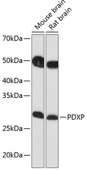
Western blot analysis of various lysates using PDXP Rabbit pAb (CAB17455) at 1:1000 dilution. Secondary antibody: HRP-conjugated Goat anti-Rabbit IgG (H+L) (CABS014) at 1:10000 dilution. Lysates/proteins: 25μg per lane. Blocking buffer: 3% nonfat dry milk in TBST. Detection: ECL Basic Kit (AbGn00020). Exposure time: 10s.
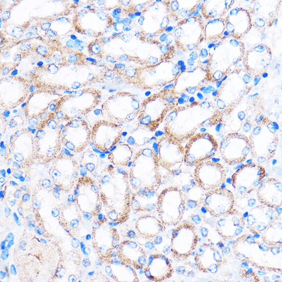
Immunohistochemistry analysis of paraffin-embedded Mouse kidney using PDXP Rabbit pAb (CAB17455) at dilution of 1:100 (40x lens). Microwave antigen retrieval performed with 0.01M PBS Buffer (pH 7.2) prior to IHC staining.
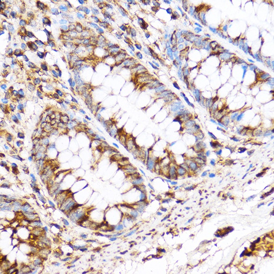
Immunohistochemistry analysis of paraffin-embedded Human colon using PDXP Rabbit pAb (CAB17455) at dilution of 1:100 (40x lens). Microwave antigen retrieval performed with 0.01M PBS Buffer (pH 7.2) prior to IHC staining.
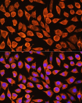
Immunofluorescence analysis of L929 cells using PDXP Rabbit pAb (CAB17455) at dilution of 1:100. Secondary antibody: Cy3-conjugated Goat anti-Rabbit IgG (H+L) (CABS007) at 1:500 dilution. Blue: DAPI for nuclear staining.