The PCCA Antibody (CAB9486) is a high-quality antibody developed for reliable detection and analysis of target proteins. This enzyme plays a key role in the breakdown of certain amino acids and fatty acids in the body. Raised in rabbits, this antibody is highly specific to human samples and is validated for use in Western blot applications. It binds specifically to the PCCA protein, allowing for accurate detection and analysis in a variety of cell types. Research involving the PCCA gene and its protein product is essential in understanding metabolic diseases such as propionic acidemia, a rare inherited disorder characterized by the body's inability to break down certain amino acids and fats.
This antibody is validated for use in WB, IHC-P, ELISA applications and has demonstrated reactivity against Human, Mouse, Rat samples.
Product Name:
PCCA Antibody
SKU:
CAB9486
Size:
20μL, 100μL
Reactivity:
Human, Mouse, Rat
Conjugate:
Unconjugated
Immunogen:
Recombinant protein (or fragment).This information is considered to be commercially sensitive.
Recommended starting concentration is 1 μg/mL. Please optimize the concentration based on your specific assay requirements.
Synonyms:
PCCA
Positive Sample:
Mouse pancreas, Mouse liver, Mouse brain, Mouse kidney, Rat kidney
Cellular Localization:
Mitochondrion Matrix.
Calculated MW:
80kDa
Observed MW:
80kDa
The protein encoded by this gene is the alpha subunit of the heterodimeric mitochondrial enzyme Propionyl-CoA carboxylase. PCCA encodes the biotin-binding region of this enzyme. Mutations in either PCCA or PCCB (encoding the beta subunit) lead to an enzyme deficiency resulting in propionic acidemia. Multiple transcript variants encoding different isoforms have been found for this gene.
Purification Method
Affinity purification
Gene ID
5095
RRID
AB_2770806
Buffer Information
Store at -20℃. Avoid freeze / thaw cycles. Buffer: Buffer: PBS containing 50% glycerol, preserved with proclin300 or sodium azide, pH 7.3.
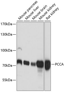
Western blot analysis of various lysates using PCCA Rabbit pAb (CAB9486) at 1:3000 dilution. Secondary antibody: HRP-conjugated Goat anti-Rabbit IgG (H+L) (CABS014) at 1:10000 dilution. Lysates/proteins: 25μg per lane. Blocking buffer: 3% nonfat dry milk in TBST. Detection: ECL Basic Kit (AbGn00020). Exposure time: 1s.
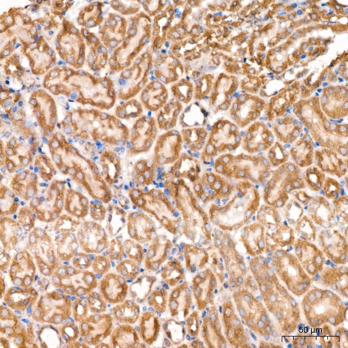
Immunohistochemistry analysis of paraffin-embedded Mouse kidney tissue using PCCA Rabbit pAb (CAB9486) at a dilution of 1:100 (40x lens). High pressure antigen retrieval was performed with 0.01 M citrate buffer (pH 6.0) prior to IHC staining.
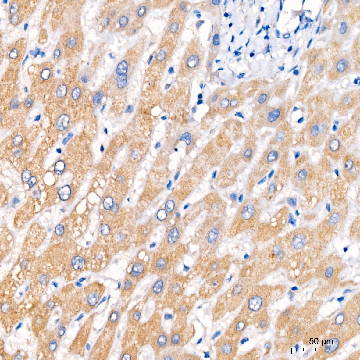
Immunohistochemistry analysis of paraffin-embedded Human liver tissue using PCCA Rabbit pAb (CAB9486) at a dilution of 1:100 (40x lens). High pressure antigen retrieval was performed with 0.01 M citrate buffer (pH 6.0) prior to IHC staining.