Description
Murine IL-2 ELISpot Pair
Assay Genie ELISpot is a highly specific immunoassay for the analysis of IL-2 production and secretion from T-cells at a single cell level in conditions closely comparable to the in-vivo environment with minimal cell manipulation. This technique is designed to determine the frequency of IL-2 producing cells under a given stimulation and the comparison of such frequency against a specific treatment or pathological state. Utilising sandwich immuno-enzyme technology, Assay Genie ELISpot assays can detect both secreted IL-2 (qualitative analysis) and single cells that produce IL-2 (quantitative analysis). Cell secreted IL-2 is captured by coated antibodies avoiding diffusion in supernatant, protease degradation or binding on soluble membrane receptors. After cell removal, the captured IL-2 is revealed by tracer antibodies and appropriate conjugates.
Murine IL-2 ELISpot Pair
The Murine IL-2 ELISPOT Pair offered by AssayGenie is a powerful tool for accurately measuring IL-2 levels in murine samples. This ELISPOT pair boasts high sensitivity and specificity, providing researchers with reliable and reproducible results for a variety of research applications.IL-2 is a key cytokine involved in regulating the immune response, particularly in T cell proliferation and activation. Monitoring IL-2 levels is vital in understanding immune system function, autoimmune disorders, and infectious diseases.
The Murine IL-2 ELISPOT Pair is an essential tool for studying these conditions and developing novel therapeutic interventions.Overall, the Murine IL-2 ELISPOT Pair from AssayGenie is a valuable asset for researchers looking to delve deeper into the role of IL-2 in murine models and investigate new avenues for treatment and diagnosis.
| Product type: | ELISpot Pairs |
| Size: | 10 x 96 Assays |
| Target species: | Murine |
| Specificity: | Recognizes natural murine IL-2 |
| Incubation: | |
| Kit content: | Assay Genie ELISpot matched antibody pairs are extensively validated and include pre-titrated capture antibody and biotinylated detection antibody. Antibodies are supplied in quantities sufficient for 10 x 96 samples. |
| Synonyms: | N/A |
| Uniprot: | P04351 |
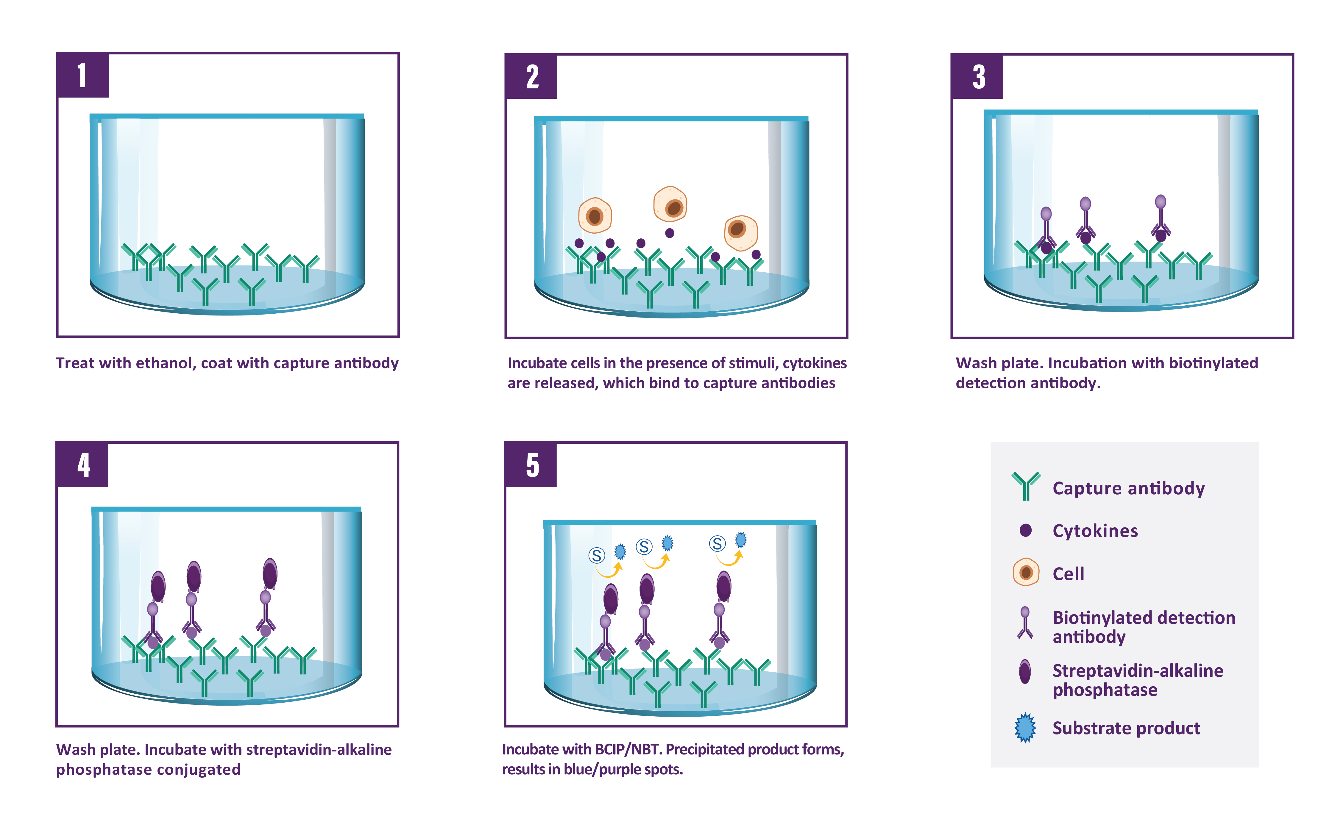
A capture antibody highly specific for IL-2 is coated to the wells of a PVDF bottomed 96 well microtitre plate either during kit manufacture or in the laboratory. The plate is then blocked to minimise any non-antibody dependent unspecific binding and washed. Cell suspension and stimulant are added and the plate incubated allowing the specific antibodies to bind any IL-2 produced. Cells are then removed by washing prior to the addition of Biotinylated detection antibodies which bind to the previously captured IL-2. Enzyme conjugated streptavidin is then added binding to the detection antibodies. Following incubation and washing, substrate is then applied to the wells resulting in coloured spots which can be quantified using appropriate analysis software or manually using a microscope.

| Step | Procedure |
| 1. | Add 100 µl of PBS 1X to every well. |
| 2. | Incubate plate at room temperature (RT) for 10 min. |
| 2. | Incubate plate at room temperature (RT) for 10 min. |
| 3. | Empty the wells by flicking the plate over a sink & gently tapping on absorbent paper. |
| 3. | Empty the wells by flicking the plate over a sink & gently tapping on absorbent paper. |
| 4. | Add 100 µl of sample, positive and negative controls cell suspension to appropriate wells providing the required concentration of cells and stimulant. |
| 5. | Cover the plate and incubate at 37°C in a CO2 incubator for an appropriate length of time (15-20 hours). Note: do not agitate or move the plate during this incubation. |
| 6. | Empty the wells and remove excess solution then add 100 µl of Wash Buffer to every well. |
| 7. | Incubate the plate at 4°C for 10 min. |
| 8. | Empty the wells as previous and wash the plate 3x with 100 µl of Wash Buffer. |
| 9. | Add 100 µl of diluted detection antibody to every well. |
| 10. | Cover the plate and incubate at RT for 1 hour 30 min. |
| 11. | Empty the wells as previous and wash the plate 3x with 100 µl of Wash Buffer. |
| 12. | Add 100 µl of diluted Streptavidin-AP conjugate to every well. |
| 13. | Cover the plate and incubate at RT for 1 hour. |
| 14. | Empty the wells and wash the plate 3x with 100 µl of Wash Buffer. |
| 15. | Peel of the plate bottom and wash both sides of the membrane 3x under running distilled water, once washing is complete remove any excess solution by repeated tapping on absorbent paper. |
| 16. | Add 100 µl of ready-to-use BCIP/NBT buffer to every well. |
| 17. | Incubate the plate for 5-15 min monitoring spot formation visually throughout the incubation period to assess sufficient colour development. |
| 18. | Empty the wells and rinse both sides of the membrane 3x under running distilled water. Completely remove any excess solution by gentle repeated tapping on absorbent paper. |
| 19. | Read Spots: allow the wells to dry and then read results. The frequency of the resulting coloured spots corresponding to the cytokine producing cells can be determined using an appropriate ELISpot reader and analysis software or manually using a microscope. Note: spots may become sharper after overnight incubation at 4°C in the dark. |
| UniProt Protein Function: | IL2: Produced by T-cells in response to antigenic or mitogenic stimulation, this protein is required for T-cell proliferation and other activities crucial to regulation of the immune response. Can stimulate B-cells, monocytes, lymphokine- activated killer cells, natural killer cells, and glioma cells. A chromosomal aberration involving IL2 is found in a form of T-cell acute lymphoblastic leukemia (T-ALL). Translocation t(4;16)(q26;p13) with involves TNFRSF17. Belongs to the IL-2 family. |
| UniProt Protein Details: | Protein type:Secreted, signal peptide; Secreted; Oncoprotein; Cytokine Cellular Component: cytosol; extracellular region; extracellular space Molecular Function:carbohydrate binding; cytokine activity; glycosphingolipid binding; growth factor activity; interleukin-2 receptor binding; kappa-type opioid receptor binding Biological Process: adaptive immune response; elevation of cytosolic calcium ion concentration; G-protein coupled receptor protein signaling pathway; immune response; immune system process; negative regulation of B cell apoptosis; negative regulation of heart contraction; negative regulation of inflammatory response; negative regulation of lymphocyte proliferation; negative regulation of protein amino acid phosphorylation; positive regulation of activated T cell proliferation; positive regulation of B cell proliferation; positive regulation of immunoglobulin secretion; positive regulation of interferon-gamma production; positive regulation of interleukin-17 production; positive regulation of isotype switching to IgG isotypes; positive regulation of protein amino acid phosphorylation; positive regulation of regulatory T cell differentiation; positive regulation of T cell differentiation; positive regulation of T cell proliferation; positive regulation of transcription from RNA polymerase II promoter; positive regulation of tyrosine phosphorylation of Stat5 protein; protein kinase C activation; regulation of T cell homeostatic proliferation; response to ethanol |
| UniProt Code: | P04351 |
| NCBI GenInfo Identifier: | 124326 |
| NCBI Gene ID: | 16183 |
| NCBI Accession: | P04351.1 |
| UniProt Secondary Accession: | P04351,P97945, Q791T3, |
| UniProt Related Accession: | P04351 |
| Molecular Weight: | 19,400 Da |
| NCBI Full Name: | Interleukin-2 |
| NCBI Synonym Full Names: | interleukin 2 |
| NCBI Official Symbol: | Il2‚ ‚ |
| NCBI Official Synonym Symbols: | Il-2‚ ‚ |
| NCBI Protein Information: | interleukin-2 |
| UniProt Protein Name: | Interleukin-2 |
| UniProt Synonym Protein Names: | T-cell growth factor; TCGF |
| Protein Family: | Interleukin |
| UniProt Gene Name: | Il2‚ ‚ |
| UniProt Entry Name: | IL2_MOUSE |


