Histamine is a biogenic amine and signaling molecule involved in various physiological processes, including allergic responses, inflammation, and gastric acid secretion. It acts by binding to specific histamine receptors, triggering specific cellular responses. The Assay Genie Mouse histamine ELISA kit allows for the quantitative measurement of histamine levels in mouse samples, such as serum, plasma, tissue lysates, and cell culture supernatants. It provides researchers with a sensitive and reliable method to assess histamine concentrations, enabling the investigation of histamine-related processes and their role in various physiological and pathological conditions in mouse models.
Description
Mouse Histamine ELISA Kit
Key Features
| Save Time | Pre-coated 96 well plate | |
| Quick Start | Kit includes all necessary reagents | |
| Publication Ready | Reproducible and reliable results |
Overview
| Product Name: | Mouse Histamine (HIS) ELISA Kit |
| Product Code: | MOEB2524 |
| Size: | 96 Assays |
| Alias: | Histamine, HIS |
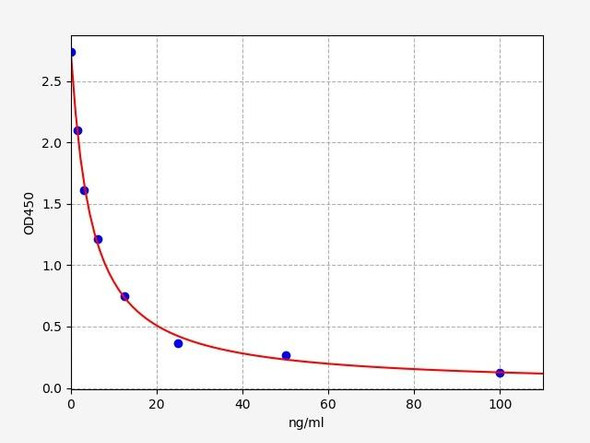
| Detection Method: | Competitive |
| Reactivity: | General |
| Range: | 7.8-500 ng/mL |
| Storage: | 4°C for 6 months |
| Note: | For Research Use Only |
Kit Components
| Component | Quantity | Storage |
| ELISA Microplate (Dismountable) | 8x12 strips | 4°C for 6 months |
| Lyophilized Standard | 2 | 4°C/ -20°C |
| Sample/Standard Dlution Buffer | 20ml | 4°C |
| Biotin-labeled Antibody (Concentrated) | 120ul | 4°C (Protection from light) |
| Antibody Dilution Buffer | 10ml | 4°C |
| HRP-Streptavidin Conjugate (SABC) | 120ul | 4°C (Protect from light) |
| SABC Dilution Buffer | 10ml | 4°C |
| TMB Substrate | 10ml | 4°C (Protection from light) |
| Stop Solution | 10ml | 4°C |
| Wash Buffer (25X) | 30ml | 4°C |
| Plate Sealer | 5 | - |
Other materials required:
- Microplate reader with 450 nm wavelength filter
- Multichannel Pipette, Pipette, microcentrifuge tubes and disposable pipette tips
- Incubator
- Deionized or distilled water
- Absorbent paper
- Buffer resevoir
Protocol
*Note: Protocols are specific to each batch/lot. For the correct instructions please follow the protocol included in your kit.
Equilibrate the TMB substrate for at least 30 min at 37°C beforeuse. When diluting samples and reagents, they must be mixed completely andevenly. It is recommended to plot a standard curve for each test.
| Step | Procedure |
| 1. | Add 50µL of Standard, Blank, or Sample per well. The blank well is added with Sample diluent. Solutions are added to the bottom of micro ELISA plate well, avoid inside wall touching and foaming as possible. |
| 2. | Immediately add 50µL of Detection Reagent A working solution to each well. Cover with the Plate sealer. Gently tap the plate to ensure thorough mixing. Incubate for 1 hour at 37°C. Note: if Detection Reagent A appears cloudy warm to room temperature until solution is uniform. |
| 3. | Aspirate each well and wash, repeating the process three times. Wash by filling each well with Wash Buffer (approximately 400µL) (a squirt bottle, multi-channel pipette, manifold dispenser or automated washer are needed) and let it sit in the well for 1-2 minutes. Complete removal of liquid at each step is essential. After the last wash, completely remove remaining Wash Buffer by aspirating or decanting. Invert the plate and pat it against thick clean absorbent paper. |
| 4. | Add 100µL of Detection Reagent B working solution to each well. Cover with the Plate sealer. Incubate for 45 minutes at 37°C. |
| 5. | Repeat the wash process for five times as conducted in step 3. |
| 6. | Add 90µL of Substrate Solution to each well. Cover with a new Plate sealer and incubate for 10-20 minutes at 37°C. Protect the plate from light. The reaction time can be shortened or extended according to the actual color change, but this should not exceed more than 30 minutes. When apparent gradient appears in standard wells, user should terminate the reaction. |
| 7. | Add 50µL of Stop Solution to each well. If color change does not appear uniform, gently tap the plate to ensure thorough mixing. |
| 8. | Determine the optical density (OD value) of each well at once, using amicro-plate reader set to 450 nm. User should open the micro-plate reader in advance, preheat the instrument, and set the testing parameters. |
| 9. | After the experiment, store all reagents according to the specified storage temperature respectively until their expiry. |
Sample Preparation
When carrying out an ELISA assay it is important to prepare your samples in order to achieve the best possible results. Below we have a list of procedures for the preparation of samples for different sample types.
| Sample Type | Protocol |
| Serum | If using serum separator tubes, allow samples to clot for 30 minutes at room temperature. Centrifuge for 10 minutes at 1,000x g. Collect the serum fraction and assay promptly or aliquot and store the samples at -80°C. Avoid multiple freeze-thaw cycles. If serum separator tubes are not being used, allow samples to clot overnight at 2-8°C. Centrifuge for 10 minutes at 1,000x g. Remove serum and assay promptly or aliquot and store the samples at -80°C. Avoid multiple freeze-thaw cycles. |
| Plasma | Collect plasma using EDTA or heparin as an anticoagulant. Centrifuge samples at 4°C for 15 mins at 1000 × g within 30 mins of collection. Collect the plasma fraction and assay promptly or aliquot and store the samples at -80°C. Avoid multiple freeze-thaw cycles. Note: Over haemolysed samples are not suitable for use with this kit. |
| Urine & Cerebrospinal Fluid | Collect the urine (mid-stream) in a sterile container, centrifuge for 20 mins at 2000-3000 rpm. Remove supernatant and assay immediately. If any precipitation is detected, repeat the centrifugation step. A similar protocol can be used for cerebrospinal fluid. |
| Cell culture supernatant | Collect the cell culture media by pipette, followed by centrifugation at 4°C for 20 mins at 1500 rpm. Collect the clear supernatant and assay immediately. |
| Cell lysates | Solubilize cells in lysis buffer and allow to sit on ice for 30 minutes. Centrifuge tubes at 14,000 x g for 5 minutes to remove insoluble material. Aliquot the supernatant into a new tube and discard the remaining whole cell extract. Quantify total protein concentration using a total protein assay. Assay immediately or aliquot and store at ≤ -20 °C. |
| Tissue homogenates | The preparation of tissue homogenates will vary depending upon tissue type. Rinse tissue with 1X PBS to remove excess blood & homogenize in 20ml of 1X PBS (including protease inhibitors) and store overnight at ≤ -20°C. Two freeze-thaw cycles are required to break the cell membranes. To further disrupt the cell membranes you can sonicate the samples. Centrifuge homogenates for 5 mins at 5000xg. Remove the supernatant and assay immediately or aliquot and store at -20°C or -80°C. |
| Tissue lysates | Rinse tissue with PBS, cut into 1-2 mm pieces, and homogenize with a tissue homogenizer in PBS. Add an equal volume of RIPA buffer containing protease inhibitors and lyse tissues at room temperature for 30 minutes with gentle agitation. Centrifuge to remove debris. Quantify total protein concentration using a total protein assay. Assay immediately or aliquot and store at ≤ -20 °C |
| Breast Milk | Collect milk samples and centrifuge at 10,000 x g for 60 min at 4°C. Aliquot the supernatant and assay. For long term use, store samples at -80°C. Minimize freeze/thaw cycles. |
Histamine Background
What is Histamine?
Histamine is a biogenic amine that serves as a vital neurotransmitter and signaling molecule in the human body. It is involved in various physiological processes and plays a crucial role in the immune response. Histamine is synthesized and stored in mast cells and basophils, which are types of white blood cells found in tissues throughout the body. When histamine is released, it can trigger a range of effects, including vasodilation, increased vascular permeability, smooth muscle contraction, and stimulation of gastric acid secretion.
Histamine Structure
Histamine has a simple chemical structure consisting of an imidazole ring connected to an ethylamine side chain. The imidazole ring is crucial for its biological activity, as it allows histamine to bind to specific receptors, namely H1, H2, H3, and H4 receptors. These receptors are found on the surface of various cells and tissues throughout the body, enabling histamine to exert its diverse physiological effects.
Structure of Histamine. Source: PubChem
Histamine Function
Histamine performs a wide range of functions in the body. It acts as a neurotransmitter in the central nervous system, playing a role in regulating sleep-wake cycles, appetite, and cognitive functions. In peripheral tissues, histamine functions as a potent mediator of allergic and inflammatory responses. It is involved in the immediate hypersensitivity reaction, contributing to the symptoms of allergies, such as itching, sneezing, and nasal congestion. Histamine also plays a crucial role in gastric acid secretion, regulating stomach acid levels and aiding in the digestion process.
Histamine Synthesis
Histamine is synthesized in the body through a two-step enzymatic process. The first step involves the decarboxylation of the amino acid histidine by the enzyme histidine decarboxylase (HDC), which removes a carboxyl group and forms histamine. This reaction takes place primarily in mast cells and basophils. The second step involves the enzymatic methylation of histamine by histamine N-methyltransferase (HNMT) in various tissues, including the liver, kidneys, and central nervous system, to form N-methylhistamine. These enzymes play a crucial role in regulating histamine levels and its subsequent physiological effects.
Histamine Receptors
There are four known types of histamine receptors: H1, H2, H3, and H4. Each receptor subtype is associated with different physiological functions and is found in different locations within the body. When histamine binds to its respective receptor, it triggers a series of intracellular signaling pathways that lead to specific cellular responses. For example, activation of H1 receptors can cause smooth muscle contraction, increased vascular permeability, and allergic symptoms, while activation of H2 receptors stimulates gastric acid secretion. H3 and H4 receptors are mainly found in the central nervous system and immune cells, respectively, and are involved in regulating neurotransmitter release and modulating immune and inflammatory responses.
Histamine Processing
Once histamine is synthesized, it is stored in vesicles within mast cells and basophils. Upon activation by various triggers like allergens, physical trauma, or immune responses, these cells release histamine into the surrounding tissues. Histamine can then bind to its specific receptors (H1, H2, H3, and H4) located on various cells and tissues throughout the body, initiating specific physiological responses. After exerting its effects, histamine undergoes enzymatic degradation. The primary enzyme involved in histamine metabolism is diamine oxidase (DAO), which breaks down histamine into imidazole acetic acid, reducing its biological activity. Another enzyme, histamine N-methyltransferase (HNMT), also plays a role in metabolizing histamine by methylating it to form N-methylhistamine. These enzymatic processes help regulate histamine levels and prevent excessive histamine accumulation in the body.
Histamine Clinical Significance
Histamine's clinical significance is vast, with both therapeutic and pathological implications. Excessive histamine release or impaired histamine metabolism can lead to various allergic disorders, including allergic rhinitis, asthma, and urticaria (hives). Antihistamines are commonly used to alleviate these symptoms by blocking histamine receptors. Additionally, histamine is involved in gastric acid secretion, and abnormalities in histamine signaling can contribute to gastrointestinal disorders like peptic ulcers. Understanding the role of histamine and its receptors has led to the development of medications targeting specific receptor subtypes, providing effective treatments for conditions like gastric acid-related diseases and allergic reactions.
Histamine Intolerance
Histamine intolerance is a condition characterized by the body's inability to properly metabolize histamine. It occurs due to a deficiency or dysfunction of enzymes responsible for histamine breakdown. When histamine accumulates, it can cause symptoms similar to allergies, such as headaches, itching, digestive issues, and nasal congestion. Diagnosis involves evaluating symptoms and dietary elimination trials. Treatment focuses on reducing histamine levels by following a low-histamine diet and, in some cases, using supplements. Histamine intolerance is different from a true allergy, as it involves impaired histamine processing rather than an immune response.
Mouse Histamine ELISA Kit FAQs
Q: What is the purpose of the mouse histamine ELISA kit?
The mouse histamine ELISA kit is specifically designed for the quantitative detection and measurement of histamine levels in mouse samples. It provides a sensitive and reliable method to assess histamine concentrations, allowing researchers to investigate histamine-related processes, allergic responses, inflammation, and other physiological and pathological conditions in mouse models.
Q: What type of samples can be used with the mouse histamine ELISA kit?
The mouse histamine ELISA kit is compatible with various sample types, including mouse serum, plasma, tissue lysates, and cell culture supernatants. It allows researchers to analyze different biological matrices and provides flexibility in sample selection based on the specific experimental needs.
Q: Where can I find additional technical support or assistance with the Mouse Histamine ELISA kit?
For any technical inquiries or assistance regarding the Mouse Histamine ELISA kit, you can reach out to our team. They will be available to answer your questions and provide the necessary guidance to ensure a successful experiment.
Related Products

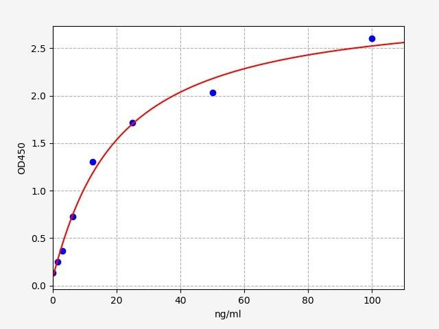
| Mouse Diamine Oxidase ELISA Kit | |
|---|---|
| ELISA TYPE: | Sandwich ELISA, Double Antibody |
| SENSITIVITY: | 0.938ng/ml |
| RANGE: | 1.563-100ng/ml |

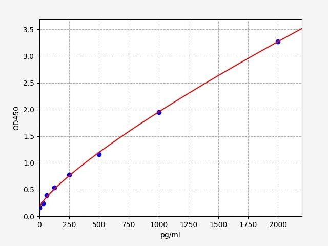
| Human HNMT ELISA Kit | |
|---|---|
| ELISA TYPE: | Sandwich ELISA, Double Antibody |
| SENSITIVITY: | 18.75pg/ml |
| RANGE: | 31.25-2000pg/ml |

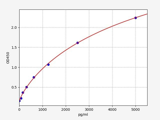
| Mouse Tryptase ELISA Kit | |
|---|---|
| ELISA TYPE: | Sandwich ELISA, Double Antibody |
| SENSITIVITY: | 46.875pg/ml |
| RANGE: | 78.125-5000pg/ml |


