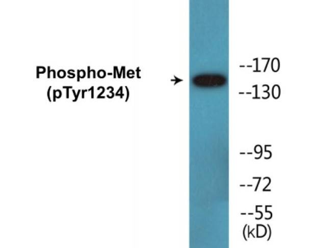
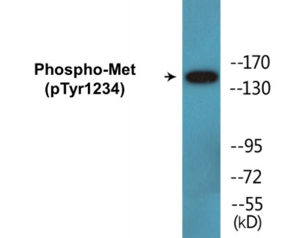
Description
Met (Phospho-Tyr1234) Cell-Based ELISA Kit
The Met (Phospho-Tyr1234) Cell-Based ELISA Kit is a convenient, lysate- free, high throughput and sensitive assay kit that can monitor Met phosphorylation and expression profile in cells. The kit can be used for measuring the relative amounts of phosphorylated Met in cultured cells as well as screening for the effects that various treatments, inhibitors (ie. siRNA or chemicals), or activators have on Met phosphorylation.
How does our Met (Phospho-Tyr1234) Fluorometric Cell-Based ELISA Kit work?
Qualitative determination of Met (Phospho-Tyr1234) concentration is achieved by an indirect ELISA format. In essence, Met (Phospho-Tyr1234) is captured by Met (Phospho-Tyr1234)-specific primary (1°) antibodies while Dye 1-conjugated and Dye 2-conjugated secondary (2°) antibodies bind the Fc region of the 1° antibody. Through this binding, the dye conjugated to the 2° antibody can emit light at a certain wavelength given proper excitation, hence allowing for a fluorometric detection method. Due to the qualitative nature of the Cell-Based ELISA, multiple normalization methods are needed:
| 1. | A monoclonal antibody specific for human GAPDH is included to serve as an internal positive control in normalizing the target RFU values. |
| 2. | An antibody against the nonphosphorylated counterpart of Met (Phospho-Tyr1234) is also provided for normalization purposes. The RFU values obtained for non-phosphorylated Met can be used to normalize the RFU value for phosphorylated Met. |
Met (Phospho-Tyr1234) Fluorometric Cell-Based ELISA Kit -Information
| Product Name: | Met (Phospho-Tyr1234) Fluorometric Cell-Based ELISA Kit |
| Product Code/SKU: | FBCAB00071 |
| Description: | The Met (Phospho-Tyr1234) Fluorometric Cell-Based Phospho ELISA Kit is a convenient, lysate-free, high throughput and sensitive assay kit that can monitor Met (Phospho-Tyr1234) protein phosphorylation and expression profile in cells. The kit can be used for measuring the relative amounts of phosphorylated Met (Phospho-Tyr1234) in cultured cells as well as screening for the effects that various treatments, inhibitors (ie. siRNA or chemicals, or activators have on MET phosphorylation. |
| Dynamic Range: | > 5000 Cells |
| Detection Method: | Fluorometric |
| Storage/Stability: | 4°C/6 Months |
| Reactivity: | Human, Mouse, Rat |
| Assay Type: | Cell-Based ELISA |
| Database Links: | Gene ID: 4233, UniProt ID: P08581, OMIM #: 114550/164860/605074/611015, Unigene #: Hs.132966 |
| Format: | Two 96-Well Plates |
| NCBI Gene Symbol: | MET |
| Sub Type: | Phospho |
| Target Name: | Phospho-Met (Tyr1234) |
Kit Principle
Figure: Schematic representation of Assay Genie Cell-Based Fluorometric ELISA principle
Kit components | Quantity |
| 96-Well Black Cell CultureClear-Bottom Microplate | 2 plates |
| 10X TBS | 24 ml |
| Quenching Buffer | 24 ml |
| Blocking Buffer | 50 ml |
| 15X Wash Buffer | 50 ml |
| Primary Antibody Diluent | 12 ml |
| 100x Anti-Phospho Target Antibody | 60 µl |
| 100x Anti-Target Antibody | 60 µl |
| Anti-GAPDH Antibody | 110 µl |
| Dye-1 Conjugated Anti-Rabbit IgG Antibody | 6 ml |
| Dye-2 Conjugated Anti-Mouse IgG Antibody | 6 ml |
| Adhesive Plate Seals | 2 seals |
Additional equipment and materials required
The following materials and/or equipment are NOT provided in this kit but are necessary to successfully conduct the experiment:
- Fluorescent plate reader with two channels at Ex/Em: 651/667 and 495/521
- Micropipettes capable of measuring volumes from 1 µl to 1 ml
- Deionized or sterile water (ddH2O)
- 37% formaldehyde (Sigma Cat# F-8775) or formaldehyde from other sources
- Squirt bottle, manifold dispenser, multichannel pipette reservoir or automated microplate washer
- Graph paper or computer software capable of generating or displaying logarithmic functions
- Absorbent papers or vacuum aspirator
- Test tubes or microfuge tubes capable of storing ≥1 ml
- Poly-L-Lysine (Sigma Cat# P4832 for suspension cells)
- Orbital shaker (optional)
Kit Protocol
This is a summarized version of the kit protocol. Please view the technical manual of this kit for information on sample preparation, reagent preparation and plate lay out.
| 1. | Seed 200 µl of desired cell concentration in culture medium into each well of the 96-well plates. For suspension cells and loosely attached cells, coat the plates with 100 µl of 10 µg/ml Poly-L-Lysine (not included) to each well of a 96-well plate for 30 minutes at 37°C prior to adding cells. |
| 2. | Incubate the cells for overnight at 37°C, 5% CO2. |
| 3. | Treat the cells as desired. |
| 4. | Remove the cell culture medium and rinse with 200 µl of 1x TBS, twice. |
| 5. | Fix the cells by incubating with 100 µl of Fixing Solution for 20 minutes at room temperature. The 4% formaldehyde is used for adherent cells and 8% formaldehyde is used for suspension cells and loosely attached cells. |
| 6. | Remove the Fixing Solution and wash the plate 3 times with 200 µl 1x Wash Buffer for 3 minutes. The plate can be stored at 4°C for a week. |
| 7. | Add 100 µl of Quenching Buffer and incubate for 20 minutes at room temperature. |
| 8. | Wash the plate 3 times with 1x Wash Buffer for 3 minutes each time. |
| 9. | Dispense 200 µl of Blocking Buffer and incubate for 1 hour at room temperature. |
| 10. | Wash 3 times with 200 µl of 1x Wash Buffer for 3 minutes each time. |
| 11. | Add 50 µl of Primary Antibody Mixture P to corresponding wells for Met (Phospho-Tyr1234) detection. Add 50 µl of Primary Antibody Mixture NP to the corresponding wells for total Met detection. Cover the plate with parafilm and incubate for 16 hours (overnight) at 4°C. If the target expression is known to be high, incubate for 2 hours at room temperature. |
| 12. | Wash 3 times with 200 µl of 1x Wash Buffer for 3 minutes each time. |
| 13. | Add 50 ul of Secondary Antibody Mixture to corresponding wells and incubate for 1.5 hours at room temperature in the dark. |
| 14. | Wash 3 times with 200 µl of 1x Wash Buffer for 3 minutes each time. |
| 15. | Read the plate(s) at Ex/Em: 651/667 (Dye 1) and 495/521 (Dye 2). Shield plates from direct light exposure. |
| 16. | Wash 3 times with 200 µl of 1x Wash Buffer for 5 minutes each time. |
Met (Phospho-Tyr1234) - Protein Information
| UniProt Protein Function: | Receptor tyrosine kinase that transduces signals from the extracellular matrix into the cytoplasm by binding to hepatocyte growth factor/HGF ligand. Regulates many physiological processes including proliferation, scattering, morphogenesis and survival. Ligand binding at the cell surface induces autophosphorylation of MET on its intracellular domain that provides docking sites for downstream signaling molecules. Following activation by ligand, interacts with the PI3-kinase subunit PIK3R1, PLCG1, SRC, GRB2, STAT3 or the adapter GAB1. Recruitment of these downstream effectors by MET leads to the activation of several signaling cascades including the RAS-ERK, PI3 kinase-AKT, or PLCgamma-PKC. The RAS-ERK activation is associated with the morphogenetic effects while PI3K/AKT coordinates prosurvival effects. During embryonic development, MET signaling plays a role in gastrulation, development and migration of muscles and neuronal precursors, angiogenesis and kidney formation. In adults, participates in wound healing as well as organ regeneration and tissue remodeling. Promotes also differentiation and proliferation of hematopoietic cells. May regulate cortical bone osteogenesis. |
| NCBI Summary: | This gene encodes a member of the receptor tyrosine kinase family of proteins and the product of the proto-oncogene MET. The encoded preproprotein is proteolytically processed to generate alpha and beta subunits that are linked via disulfide bonds to form the mature receptor. Further processing of the beta subunit results in the formation of the M10 peptide, which has been shown to reduce lung fibrosis. Binding of its ligand, hepatocyte growth factor, induces dimerization and activation of the receptor, which plays a role in cellular survival, embryogenesis, and cellular migration and invasion. Mutations in this gene are associated with papillary renal cell carcinoma, hepatocellular carcinoma, and various head and neck cancers. Amplification and overexpression of this gene are also associated with multiple human cancers. [provided by RefSeq, May 2016] |
| UniProt Code: | P08581 |
| NCBI GenInfo Identifier: | 251757497 |
| NCBI Gene ID: | 4233 |
| NCBI Accession: | P08581.4 |
| UniProt Secondary Accession: | P08581,O60366, Q12875, Q9UDX7, Q9UPL8, A1L467, B5A932 E7EQ94, |
| UniProt Related Accession: | P08581 |
| Molecular Weight: | 85,745 Da |
| NCBI Full Name: | Hepatocyte growth factor receptor |
| NCBI Synonym Full Names: | MET proto-oncogene, receptor tyrosine kinase |
| NCBI Official Symbol: | MET |
| NCBI Official Synonym Symbols: | HGFR; AUTS9; RCCP2; c-Met; DFNB97 |
| NCBI Protein Information: | hepatocyte growth factor receptor |
| UniProt Protein Name: | Hepatocyte growth factor receptor |
| UniProt Synonym Protein Names: | HGF/SF receptor; Proto-oncogene c-Met; Scatter factor receptor; SF receptor; Tyrosine-protein kinase Met |
| Protein Family: | C-methyltransferase |
| UniProt Gene Name: | MET |


