The MAP1A Polyclonal Antibody (PACO56884) is a valuable tool for researchers studying the MAP1A protein, which plays a key role in microtubule stability and neuronal development. This polyclonal antibody, generated in rabbits, exhibits high specificity and sensitivity for detecting MAP1A in human samples, making it an ideal choice for Western blot applications.MAP1A is a critical regulator of microtubule dynamics in neurons, influencing processes such as cell migration, axon guidance, and synaptic plasticity.
Dysregulation of MAP1A has been linked to neurodegenerative diseases, making it an important target for further investigation in the field of neuroscience. The MAP1A Polyclonal Antibody enables accurate detection and quantification of MAP1A levels in various cell types, providing valuable insights into its functions and potential implications for neurological disorders.
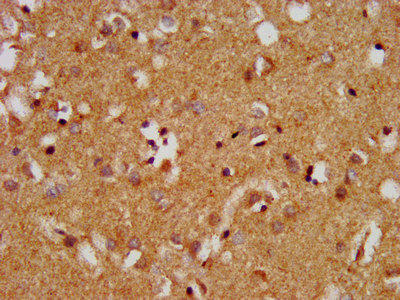
IHC image of PACO56884 diluted at 1:400 and staining in paraffin-embedded human brain tissue performed on a Leica BondTM system. After dewaxing and hydration, antigen retrieval was mediated by high pressure in a citrate buffer (pH 6.0). Section was blocked with 10% normal goat serum 30min at RT. Then primary antibody (1% BSA) was incubated at 4°C overnight. The primary is detected by a biotinylated secondary antibody and visualized using an HRP conjugated SP system.
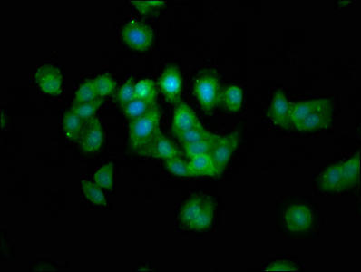
Immunofluorescence staining of HepG2 cells with PACO56884 at 1:133, counter-stained with DAPI. The cells were fixed in 4% formaldehyde, permeabilized using 0.2% Triton X-100 and blocked in 10% normal Goat Serum. The cells were then incubated with the antibody overnight at 4°C. The secondary antibody was Alexa Fluor 488-congugated AffiniPure Goat Anti-Rabbit IgG(H+L).
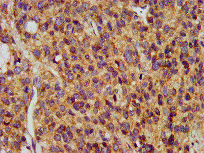
IHC image of PACO56884 diluted at 1:400 and staining in paraffin-embedded human glioma performed on a Leica BondTM system. After dewaxing and hydration, antigen retrieval was mediated by high pressure in a citrate buffer (pH 6.0). Section was blocked with 10% normal goat serum 30min at RT. Then primary antibody (1% BSA) was incubated at 4°C overnight. The primary is detected by a biotinylated secondary antibody and visualized using an HRP conjugated SP system.
Background:
Structural protein involved in the filamentous cross-bridging between microtubules and other skeletal elements.
Synonyms:
Microtubule-associated protein 1A (MAP-1A) (Proliferation-related protein p80) [Cleaved into: MAP1A heavy chain; MAP1 light chain LC2], MAP1A, MAP1L
UniProt Protein Function:
MAP1A: structural protein involved in the filamentous cross- bridging between microtubules and other skeletal elements. Various serine residues may be phosphorylated by PKA.Protein type: Motility/polarity/chemotaxis; CytoskeletalChromosomal Location of Human Ortholog: 15q15.3Cellular Component: microtubule; microtubule associated complex; cytosolMolecular Function: protein binding; microtubule binding; structural molecule activityBiological Process: sensory perception of sound; microtubule cytoskeleton organization and biogenesis
UniProt Protein Details:
NCBI Summary:
This gene encodes a protein that belongs to the microtubule-associated protein family. The proteins of this family are thought to be involved in microtubule assembly, which is an essential step in neurogenesis. The product of this gene is a precursor polypeptide that presumably undergoes proteolytic processing to generate the final MAP1A heavy chain and LC2 light chain. Expression of this gene is almost exclusively in the brain. Studies of the rat microtubule-associated protein 1A gene suggested a role in early events of spinal cord development. [provided by RefSeq, Jul 2008]


. Section was blocked with 10% normal goat serum 30min at RT. Then primary antibody (1% BSA) was incubated at 4°C overnight. The primary is detected by a Goat anti-rabbit IgG labeled by HRP and visualized using 0.05% DAB.")
.")

.")
.")
. Section was blocked with 10% normal goat serum 30min at RT. Then primary antibody (1% BSA) was incubated at 4°C overnight. The primary is detected by a Goat anti-rabbit polymer IgG labeled by HRP and visualized using 0.05% DAB.")