The KRT18 Antibody (PACO60745) is a polyclonal antibody specifically designed for research involving Keratin 18 (KRT18), a type I keratin protein that is primarily expressed in simple epithelial tissues. This antibody, generated in rabbits, is highly specific and reactive with human samples, making it an excellent tool for studying the expression and localization of KRT18 in various cell types.KRT18 is a key component of the cytoskeleton and is involved in maintaining the structural integrity of epithelial cells. Abnormal expression of KRT18 has been linked to various diseases, including liver disease, inflammatory disorders, and certain types of cancer. Research using the KRT18 Antibody can provide valuable insights into the role of KRT18 in these pathological conditions and may lead to the development of targeted therapies.
Validated for use in Western blot applications, the KRT18 Antibody binds specifically to the KRT18 protein, allowing for the detection and analysis of KRT18 levels in experimental samples. This antibody is an essential tool for scientists and researchers working in the fields of cell biology, cancer research, and molecular biology. By studying KRT18 expression and function, researchers can expand our understanding of the complex mechanisms underlying various diseases and potentially identify new therapeutic targets.
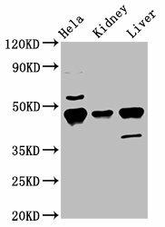
Western Blot. Positive WB detected in: Hela whole cell lysate, Mouse kidney tissue, Mouse liver tissue. All lanes: Krt18 antibody at 5.5µg/ml. Secondary. Goat polyclonal to rabbit IgG at 1/50000 dilution. Predicted band size: 48 kDa. Observed band size: 48 kDa.
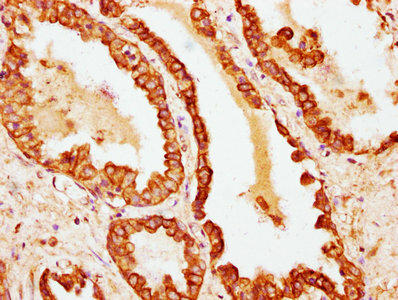
IHC image of PACO60745 diluted at 1:100 and staining in paraffin-embedded human prostate cancer performed on a Leica BondTM system. After dewaxing and hydration, antigen retrieval was mediated by high pressure in a citrate buffer (pH 6.0). Section was blocked with 10% normal goat serum 30min at RT. Then primary antibody (1% BSA) was incubated at 4°C overnight. The primary is detected by a biotinylated secondary antibody and visualized using an HRP conjugated SP system.
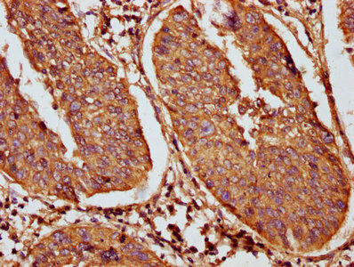
IHC image of PACO60745 diluted at 1:100 and staining in paraffin-embedded human cervical cancer performed on a Leica BondTM system. After dewaxing and hydration, antigen retrieval was mediated by high pressure in a citrate buffer (pH 6.0). Section was blocked with 10% normal goat serum 30min at RT. Then primary antibody (1% BSA) was incubated at 4°C overnight. The primary is detected by a biotinylated secondary antibody and visualized using an HRP conjugated SP system.
Background:
When phosphorylated, plays a role in filament reorganization. Involved in the delivery of mutated CFTR to the plasma membrane. Involved in the uptake of thrombin-antithrombin complexes by hepatic cells (By similarity). Together with KRT8, is involved in interleukin-6 (IL-6)-mediated barrier protection.
Synonyms:
Keratin, type I cytoskeletal 18 (Cytokeratin endo B) (Keratin D) (Cytokeratin-18) (CK-18) (Keratin-18) (K18), Krt18, Kerd Krt1-18
UniProt Protein Function:
When phosphorylated, plays a role in filament reorganization. Involved in the delivery of mutated CFTR to the plasma membrane. Involved in the uptake of thrombin-antithrombin complexes by hepatic cells. Together with KRT8, is involved in interleukin-6 (IL-6)-mediated barrier protection.


-S33 antibody . Secondary antibody: HRP Goat Anti-Rabbit IgG (H+L) at 1:10000 dilution. Lysates/proteins: 25ug per lane. Blocking buffer: 3% BSA.")


