The TXNDC9 Polyclonal Antibody (CAB19869) is a valuable tool for research involving the TXNDC9 protein, which plays a key role in redox homeostasis and protein folding in the endoplasmic reticulum. This antibody, generated in rabbits, has been rigorously validated for specificity and sensitivity in Western blot applications, making it an ideal choice for studying TXNDC9 expression and function in various cell types.TXNDC9, also known as endoplasmic reticulum protein 46 (ERp46), is involved in maintaining cellular redox balance and ensuring proper protein folding, crucial for the function of numerous proteins in the cell. Dysregulation of TXNDC9 has been linked to various diseases, including neurodegenerative disorders and cancer.
By using the TXNDC9 Polyclonal Antibody, researchers can investigate the role of TXNDC9 in disease pathogenesis and potentially identify new therapeutic targets for intervention. Overall, the TXNDC9 Polyclonal Antibody is a reliable tool for studying the function and regulation of TXNDC9 in cell biology, molecular biology, and disease research. Its high specificity and sensitivity make it a valuable asset for scientists seeking to understand the complex mechanisms underlying redox homeostasis and protein folding in health and disease.
Product Name:
[KO Validated] TXNDC9 Rabbit Polyclonal Antibody
SKU:
CAB19869
Size:
20uL, 100uL
Isotype:
IgG
Host Species:
Rabbit
Reactivity:
Human,Mouse,Rat
Immunogen:
Recombinant fusion protein containing a sequence corresponding to amino acids 1-110 of human TXNDC9 (NP_005774.2).
The protein encoded by this gene is a member of the thioredoxin family. The exact function of this protein is not known but it is associated with cell differentiation.
Purification Method:
Affinity purification
Gene ID:
10190
Storage Buffer:
Store at -20℃. Avoid freeze / thaw cycles.Buffer: PBS with 0.01% thimerosal,50% glycerol,pH7.3.
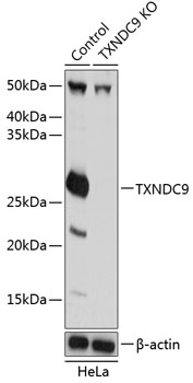
Western blot analysis of lysates from wild type (WT) and TXNDC9 knockout (KO) HeLa cells, using [KO Validated] TXNDC9 Rabbit pAb (CAB19869) at 1:1000 dilution.Secondary antibody: HRP Goat Anti-Rabbit IgG (H+L) (CABS014) at 1:10000 dilution.Lysates/proteins: 25μg per lane.Blocking buffer: 3% nonfat dry milk in TBST.Detection: ECL Basic Kit (AbGn00020).Exposure time: 1s.