The AKT2 Polyclonal Antibody (CAB18019) is a valuable tool for researchers studying the AKT2 protein, a key player in regulating cell growth and survival pathways. This antibody, produced in rabbits, is highly specific for human samples and has been validated for use in Western blot applications.AKT2, a member of the AKT family of serine/threonine protein kinases, is known for its role in various cellular processes, including cell proliferation, differentiation, and metabolism. Dysregulation of AKT2 signaling is commonly associated with cancer and other diseases, making it a promising target for therapeutic interventions.
By using the AKT2 Polyclonal Antibody, researchers can accurately detect and study the expression levels of AKT2 in different cell types and tissues. This antibody provides a reliable tool for investigating the function of AKT2 in various physiological and pathological conditions, paving the way for new insights into disease mechanisms and potential therapeutic strategies.
Product Name:
[KO Validated] AKT2 Rabbit Polyclonal Antibody
SKU:
CAB18019
Size:
20uL, 100uL
Isotype:
IgG
Host Species:
Rabbit
Reactivity:
Human,Mouse
Immunogen:
A synthetic peptide corresponding to a sequence within amino acids 381-481 of human AKT2 (NP_001617.1).
Cell membrane, Cytoplasm, Early endosome, Nucleus, Peripheral membrane protein.
Calculated MW:
56kDa
Observed MW:
60kDa
This gene is a putative oncogene encoding a protein belonging to a subfamily of serine/threonine kinases containing SH2-like (Src homology 2-like) domains, which is involved in signaling pathways. The gene serves as an oncogene in the tumorigenesis of cancer cells For example, its overexpression contributes to the malignant phenotype of a subset of human ductal pancreatic cancers. The encoded protein is a general protein kinase capable of phophorylating several known proteins, and has also been implicated in insulin signaling.
Purification Method:
Affinity purification
Gene ID:
208
Storage Buffer:
Store at -20℃. Avoid freeze / thaw cycles.Buffer: PBS with 0.01% thimerosal,50% glycerol,pH7.3.
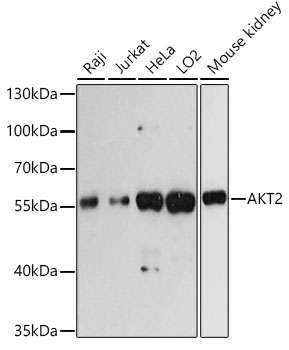
Western blot analysis of various lysates using AKT2 Rabbit pAb (CAB18019) at 1:3000 dilution.Secondary antibody: HRP Goat Anti-Rabbit IgG (H+L) (CABS014) at 1:10000 dilution.Lysates/proteins: 25μg per lane.Blocking buffer: 3% nonfat dry milk in TBST.Detection: ECL Basic Kit (AbGn00020).Exposure time: 60s.