The KCMF1 Polyclonal Antibody (PACO58524) is a valuable tool for researchers studying KCMF1, a protein involved in cell signaling and gene regulation. This antibody, produced in rabbits, has high specificity for detecting KCMF1 in human samples and is suitable for Western blot applications. By binding to the KCMF1 protein, researchers can confidently investigate its role in various cellular processes, making it an essential tool for studies in molecular biology and cancer research.KCMF1, also known as a potential tumor suppressor, is a key player in regulating cell growth and proliferation.
Its involvement in these critical pathways makes it a promising target for investigation in cancer biology and therapeutics. Understanding the function of KCMF1 can provide valuable insights into cancer development and progression, offering new opportunities for targeted therapies and personalized medicine approaches. Unlocking the mysteries of KCMF1 may lead to advancements in cancer treatment and patient care.
Antibody Name:
KCMF1 Antibody (PACO58524)
Antibody SKU:
PACO58524
Size:
50ug
Host Species:
Rabbit
Tested Applications:
ELISA, IHC
Recommended Dilutions:
ELISA:1:2000-1:10000, IHC:1:500-1:1000
Species Reactivity:
Human
Immunogen:
Recombinant Human E3 ubiquitin-protein ligase KCMF1 protein (98-226AA)
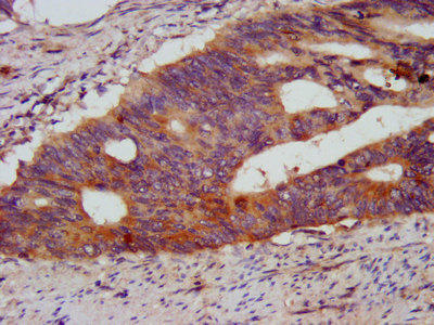
IHC image of PACO58524 diluted at 1:500 and staining in paraffin-embedded human colon cancer performed on a Leica BondTM system. After dewaxing and hydration, antigen retrieval was mediated by high pressure in a citrate buffer (pH 6.0). Section was blocked with 10% normal goat serum 30min at RT. Then primary antibody (1% BSA) was incubated at 4°C overnight. The primary is detected by a biotinylated secondary antibody and visualized using an HRP conjugated SP system.
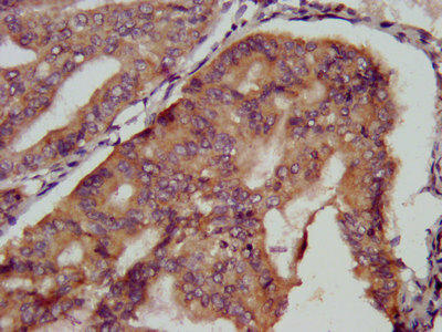
IHC image of PACO58524 diluted at 1:500 and staining in paraffin-embedded human endometrial cancer performed on a Leica BondTM system. After dewaxing and hydration, antigen retrieval was mediated by high pressure in a citrate buffer (pH 6.0). Section was blocked with 10% normal goat serum 30min at RT. Then primary antibody (1% BSA) was incubated at 4°C overnight. The primary is detected by a biotinylated secondary antibody and visualized using an HRP conjugated SP system.
Background:
Has intrinsic E3 ubiquitin ligase activity and promotes ubiquitination.


.")


.")

.")
.")