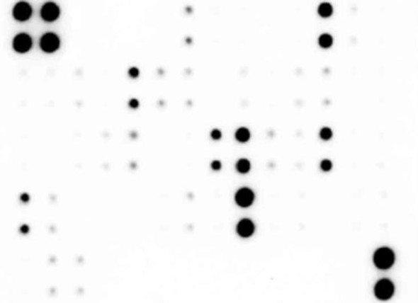
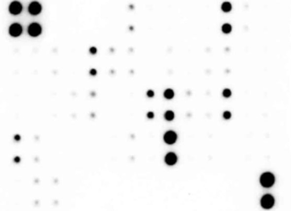
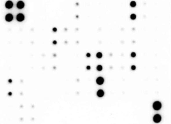
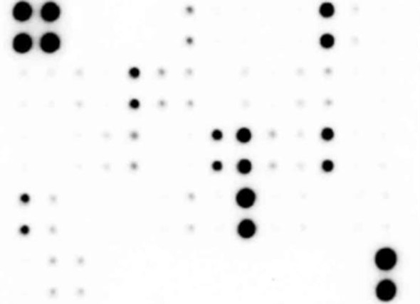
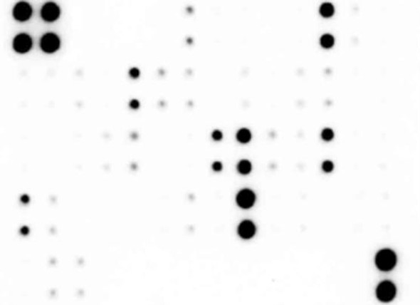
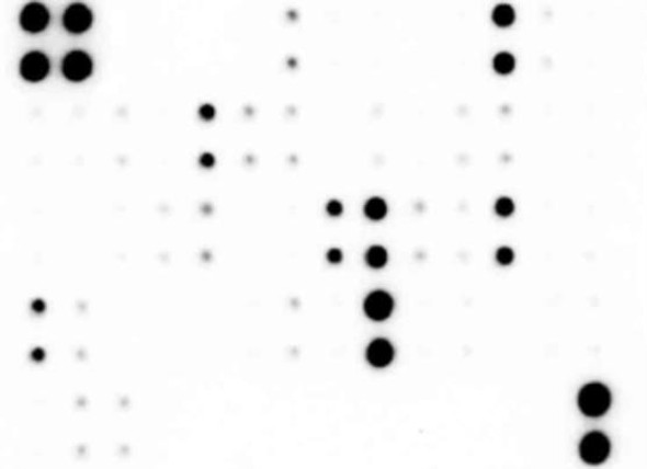
Description
Human T Cell Response Array (20 targets) (SARB0068)
The Human T Cell Response Array (SARB0068) is a comprehensive research tool designed for studying the T cell response in human samples. This array includes 20 targets related to T cell activation and function, allowing for a detailed analysis of the immune response.With a focus on T cell-related markers, this array enables researchers to identify key players in T cell signaling pathways, cytokine production, and T cell activation. By simultaneously measuring multiple targets, researchers can gain a better understanding of the complex interplay between different components of the immune response.
This array is ideal for studying T cell responses in a variety of contexts, including infectious diseases, autoimmune disorders, and cancer immunology. By providing a comprehensive view of T cell activation and function, the Human T Cell Response Array offers valuable insights for research in immunology and clinical applications.
| Product Sku: | SARB0068 |
| Size: | 2, 4, or 8 |
| Species Detected: | Human |
| Number of Targets Detected: | 20 |
| Gene Symbols: | IFNG, IL10, IL12A, IL13, IL17A, IL17F, IL18, IL2, IL21, IL22|IL23A, IL27, IL33, IL4, IL5, IL6, IL7, IL9, TGFB1, TNF |
| Compatible Sample Types: | Cell Culture Supernatants, Cell Lysates, Plasma, Serum, Tissue Lysates |
| Design Principle: | Sandwich-based |
| Method of Detection: | Chemiluminescence |
| Pathway: | |
| Quantitative/Semi-Quantitative: | Semi-Quantitative |
| Solid Support: | Membrane |
| Suggested Application(s): | Multiplexed Protein Detection; Detection of Relative Protein Expression; Detecting Patterns of Cytokine Expression; Biomarker/ Key Factor Screening; Identifying Key Factors; Confirming a Biological Process |
| Storage/Stability: | For best results, store the entire kit frozen at -20°C upon arrival. Stored frozen, the kit will be stable for at least 6 months which is the duration of the product warranty period. Once thawed, store array membranes and 1X Blocking Buffer at -20°C and all other reagents undiluted at 4°C for no more than 3 months. |
| Hover over target for more information | ||||
|---|---|---|---|---|
IL-2
| IL-4
| IL-5
| IL-6
| IL-7
|
IL-10
| IL-12 p70
| IL-13
| IFN-gamma
| TGF beta 1
|
TNF alpha
| IL-17A
| IL-9
| IL-22
| IL-17F
|
IL-18
| IL-23 p19
| IL-21
| IL-27
| IL-33 (IL-1 F11)
|
- Easy to use
- No specialized equipment needed
- Compatible with nearly any liquid sample
- Proven technology
- Highly sensitive (pg/ml)
- Sandwich ELISA specificity
- Higher density than ELISA, Western blot or bead-based multiplex
- Human T Cell Response Array C2 Membranes
- Blocking Buffer
- Wash Buffer 1
- Wash Buffer 2
- Biotinylated Detection Antibody Cocktail
- Streptavidin-Conjugated HRP
- Detection Buffer C
- Detection Buffer D
- Lysis Buffer
- 8-Well Incubation Tray
- Plastic Sheets
- Array Templates
Other Materials Required
- Pipettors, pipet tips and other common lab consumables
- Orbital shaker or oscillating rocker
- Tissue Paper, blotting paper or chromatography paper
- Adhesive tape or Saran Wrap
- Distilled or de-ionized water
- A chemiluminescent blot documentation system , X-ray Film and a suitable film processor, or another chemiluminescent detection system.
| 1. | Block membranes |
| 2. | Incubate with Sample |
| 3. | Incubate with Biotinylated Detection Antibody Cocktail |
| 4. | Incubate with HRP-Conjugated Streptavidin |
| 5. | Incubate with Detection Buffers |
| 6. | Image with chemiluminescent imaging system |
| 7. | Perform densitometry and analysis |


