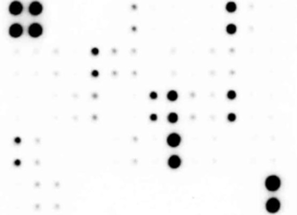
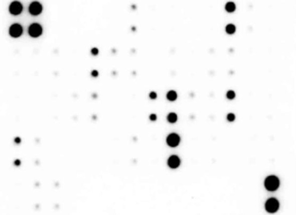
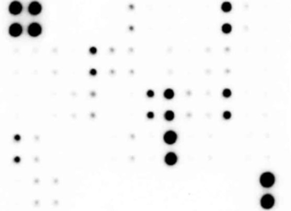
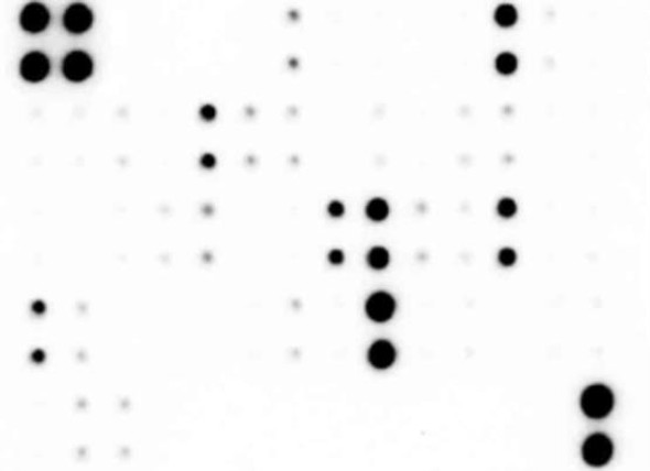
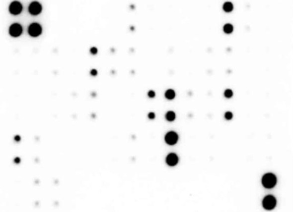
Human NF-kB Array (34 targets) (SARB0060)
- SKU:
- SARB0060
- Product Type:
- Protein Array
- Analytes:
- ASC (PYCARD)
- BCL-10
- CD40 (TNFRSF5)
- cIAP-1 (BIRC2/hIAP-2)
- cIAP-2
- c-Rel
- FADD
- Fas (TNFRSF6/Apo-1)
- IkB-alpha
- IKB-b (NFKBIB)
- IkB-epsilon (NF-kappa-BIE)
- IKK-B
- IKK-E (IKBKE)
- IKK-gamma (NEMO/FIP-3)
- IL-1 Ra (IL-1 F3)
- IL-1 R1
- IL-17 RA
- IL-1 R5 (IL-18 R alpha)
- IRAK1
- IRF5
- JNK2 (MAPK9 isoform alpha-1)
- Lymphotoxin beta R (TNF RIII)
- MyD88
- NFKB1
- nfkb2
- NGFR (TNFRSF16)
- p53
- socs6
- STING
- TLR2
- TNF RI (TNFRSF1A)
- TNF RII (TNFRSF1B)
- TRAIL R1 (TNFRSF10A/DR4)
- TRAIL R2 (TNFRSF10B/DR5)
- Reactivity:
- Human
- Applications:
- Multiplex Array
Description
Human NF-kB Array (34 targets) (SARB0060)
The Human NF-κB Array (34 targets) (SARB0060) is a powerful tool for researchers studying NF-κB signaling pathways and related processes. This array allows for the simultaneous detection and analysis of 34 NF-κB-related proteins in human samples, providing valuable insights into the activation and regulation of this key transcription factor.NF-κB is a critical regulator of immune and inflammatory responses, as well as cell survival, proliferation, and differentiation. Dysregulation of NF-κB signaling has been implicated in various diseases, including cancer, autoimmune disorders, and inflammatory conditions.
By using the Human NF-κB Array, researchers can identify changes in protein expression levels and phosphorylation status, leading to a better understanding of the molecular mechanisms underlying these diseases.This array is highly sensitive and specific, enabling the detection of low-abundance proteins and subtle changes in protein expression levels. It is easy to use, with a simple protocol that allows for rapid and reliable results. The Human NF-κB Array is an essential tool for any researcher interested in unraveling the complex role of NF-κB in health and disease.
| Product Sku: | SARB0060 |
| Size: | 2, 4, or 8 |
| Species Detected: | Human |
| Number of Targets Detected: | 34 |
| Gene Symbols: | ASC, BCL10, BIRC2, BIRC3, CD40, CIS4, FADD, FAS, FIP3, IKBB|IKBE, IKBKB, IKBKE, IL17RA, IL18R1, IL1R1, IL1RN, IRAK, IRF5, JNK2, LTBR, LYT10, MYD88, NFKB1, NGFR, REL, TLR2, TMEM173, TNFRSF10A, TNFRSF10B, TNFRSF1A, TNFRSF1B, TP53 |
| Compatible Sample Types: | Cell Culture Supernatants, Cell Lysates, Plasma, Serum, Tissue Lysates |
| Design Principle: | Sandwich-based |
| Method of Detection: | Chemiluminescence |
| Pathway: | NFkB Signaling |
| Quantitative/Semi-Quantitative: | Semi-Quantitative |
| Solid Support: | Membrane |
| Suggested Application(s): | Multiplexed Protein Detection; Detection of Relative Protein Expression; Detecting Patterns of Cytokine Expression; Biomarker/ Key Factor Screening; Identifying Key Factors; Confirming a Biological Process |
| Storage/Stability: | For best results, store the entire kit frozen at -20°C upon arrival. Stored frozen, the kit will be stable for at least 6 months which is the duration of the product warranty period. Once thawed, store array membranes and 1X Blocking Buffer at -20°C and all other reagents undiluted at 4°C for no more than 3 months. |
| Hover over target for more information | ||||
|---|---|---|---|---|
ASC (PYCARD)
| BCL-10
| CD40 (TNFRSF5)
| cIAP-1 (BIRC2/hIAP-2)
| cIAP-2
|
c-Rel
| FADD
| Fas (TNFRSF6/Apo-1)
| IkB-alpha
| IKB-b (NFKBIB)
|
IkB-epsilon (NF-kappa-BIE)
| IKK-B
| IKK-E (IKBKE)
| IKK-gamma (NEMO/FIP-3)
| IL-1 Ra (IL-1 F3)
|
IL-1 R1
| IL-17 RA
| IL-1 R5 (IL-18 R alpha)
| IRAK1
| IRF5
|
JNK2 (MAPK9 isoform alpha-1)
| Lymphotoxin beta R (TNF RIII)
| MyD88
| NFKB1
| nfkb2
|
NGFR (TNFRSF16)
| p53
| socs6
| STING
| TLR2
|
TNF RI (TNFRSF1A)
| TNF RII (TNFRSF1B)
| TRAIL R1 (TNFRSF10A/DR4)
| TRAIL R2 (TNFRSF10B/DR5)
| |
- Easy to use
- No specialized equipment needed
- Compatible with nearly any liquid sample
- Proven technology
- Highly sensitive (pg/ml)
- Sandwich ELISA specificity
- Higher density than ELISA, Western blot or bead-based multiplex
- Human NF-kB Array C2 Membranes
- Blocking Buffer
- Detection Antibody Cocktail
- 1,000X HRP-Anti-Rabbit-IgG Concentrate
- 20X Wash Buffer I Concentrate
- 20X Wash Buffer II Concentrate
- 2X Cell Lysis Buffer Concentrate
- Detection Buffer C
- Detection Buffer D
- 8-Well Incubation Tray w/ Lid
- Protease Inhibitor Cocktail
- Plastic Sheets
- Array Map Template
Other Materials Required
- Pipettors, pipet tips and other common lab consumables
- Orbital shaker or oscillating rocker
- Tissue Paper, blotting paper or chromatography paper
- Adhesive tape or plastic Wrap
- Distilled or de-ionized water
- A chemiluminescent blot documentation system: CCD camera, X-ray Film and a suitable film processor, gel documentation system, or another chemiluminescent detection system capable of imaging a western blot.
| 1. | Block membranes |
| 2. | Incubate with Sample |
| 3. | Incubate with Detection Antibody Cocktail |
| 4. | Incubate with HRP-Conjugated Streptavidin |
| 5. | Incubate with Detection Buffers |
| 6. | Image with chemiluminescent imaging system |
| 7. | Perform densitometry and analysis |

