Description
| Product Name: | Human AMH (Anti-Mullerian Hormone) ELISA Kit |
| Target: | AMH(Anti-Mullerian Hormone) |
| SKU: | AEES00739 |
| Size: | 96T |
| Reactivity: | Human |
| Sample Type: | Serum, Plasma and Other Biological Fluids; 100μL |
| Detection Method: | Sandwich ELISA |
| Assay Time: | 3.5h |
| Specificity: | This kit recognizes Human AMH in samples. No significant cross-reactivity or interference between Human AMH and analogues was observed. |
| Reproducibility: | Both intra-CV and inter-CV are < 10%. |
| Application: | This ELISA kit applies to the in vitro quantitative determination of Human AMH concentrations in serum, plasma and other biological fluids. |
| Component | Specification | Storage |
| Micro ELISA Plate(Dismountable) | 96T: 8 wells ×12 strips | -20℃, 6 months |
| Reference Standard | 96T: 2 vials | |
| Concentrated Biotinylated Detection Ab (100×) | 96T: 1 vial, 120 μL | |
| Concentrated HRP Conjugate (100×) | 96T: 1 vial, 120 μL | -20℃(Protect from light), 6 months |
| Reference Standard & Sample Diluent | 1 vial, 20 mL | 2-8°C, 6 months |
| Biotinylated Detection Ab Diluent | 1 vial, 14 mL | |
| HRP Conjugate Diluent | 1 vial, 14 mL | |
| Concentrated Wash Buffer (25×) | 1 vial, 30 mL | |
| Substrate Reagent | 1 vial, 10 mL | 2-8℃(Protect from light) |
| Stop Solution | 1 vial, 10 mL | 2-8°C |
| Plate Sealer | 5 pieces | |
| Manual | 1 copy | |
| Certificate of Analysis | 1 copy |
Other materials and equipment required:
- Microplate reader with 450 nm wavelength filter
- High-precision transfer pipette, EP tubes and disposable pipette tips
- Incubator capable of maintaining 37℃
- Deionized or distilled water
- Absorbent paper
- Loading slot
| Linearity: |
| ||||||||||||||||||||||||||||||||||||||||||
| Recovery: |
| ||||||||||||||||||||||||||||||||||||||||||
| Precision: |
| ||||||||||||||||||||||||||||||||||||||||||
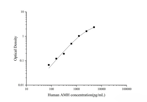
This ELISA kit uses the Sandwich-ELISA principle. The micro ELISA plate provided in this kit has been pre-coated with an antibody specific to Human AMH. Standards or samples are added to the micro ELISA plate wells and combined with the specific antibody. Then a biotinylated detection antibody specific for Human AMH and Avidin-Horseradish Peroxidase (HRP) conjugate are added successively to each micro plate well and incubated. Free components are washed away. The substrate solution is added to each well. Only those wells that contain Human AMH, biotinylated detection antibody and Avidin-HRP conjugate will appear blue in color. The enzyme-substrate reaction is terminated by the addition of stop solution and the color turns yellow. The optical density (OD) is measured spectrophotometrically at a wavelength of 450 nm ± 2 nm. The OD value is proportional to the concentration of Human AMH. You can calculate the concentration of Human AMH in the samples by comparing the OD of the samples to the standard curve.
| Step 1: | Bring all reagents to room temperature (18-25℃) before use. If the kit will not be used up in one assay, please only take out the necessary strips and reagents for present experiment, and store the remaining strips and reagents at required condition. |
| Step 2: | Wash Buffer: Dilute 30 mL of Concentrated Wash Buffer with 720 mL of deionized or distilled water to prepare 750 mL of Wash Buffer. Note: if crystals have formed in the concentrate, warm it in a 40℃ water bath and mix it gently until the crystals have completely dissolved. |
| Step 3: | Standard working solution: Centrifuge the standard at 10,000×g for 1 min. Add 1 mL of Reference Standard & Sample Diluent, let it stand for 10 min and invert it gently several times. After it dissolves fully, mix it thoroughly with a pipette. This reconstitution produces a working solution of 5000 pg/mL (or add 1 mL of Reference Standard & Sample Diluent, let it stand for 1-2 min and then mix it thoroughly with a vortex meter of low speed. Bubbles generated during vortex could be removed by centrifuging at a relatively low speed). Then make serial dilutions as needed. The recommended dilution gradient is as follows: 5000, 2500, 1250, 625, 312.5, 156.25, 78.13, 0 pg/mL. Dilution method: Take 7 EP tubes, add 500 μL of Reference Standard & Sample Diluent to each tube. Pipette 500 μL of the 5000 pg/mL working solution to the first tube and mix up to produce a 2500 pg/mL working solution. Pipette 500 μL of the solution from the former tube into the latter one according to this step. Note: the last tube is regarded as a blank. Don’t pipette solution into it from the former tube. Gradient diluted standard working solution should be prepared just before use. |
| Step 4: | Biotinylated Detection Ab working solution: Calculate the required amount before the experiment (100 μL/well). In preparation, slightly more than calculated should be prepared. Centrifuge the Concentrated Biotinylated Detection Ab at 800×g for 1 min, then dilute the 100× Concentrated Biotinylated Detection Ab to 1× working solution with Biotinylated Detection Ab Diluent (Concentrated Biotinylated Detection Ab: Biotinylated Detection Ab Diluent = 1:99). The working solution should be prepared just before use. |
| Step 5: | HRP Conjugate working solution: HRP Conjugate is HRP conjugated avidin. Calculate the required amount before the experiment (100 μL/well). In preparation, slightly more than calculated should be prepared. Centrifuge the Concentrated HRP Conjugate at 800×g for 1 min, then dilute the 100× Concentrated HRP Conjugate to 1× working solution with HRP Conjugate Diluent (Concentrated HRP Conjugate: HRP Conjugate Diluent = 1:99). The working solution should be prepared just before use. |
| Serum: | Allow samples to clot for 1 hour at room temperature or overnight at 2-8℃ before centrifugation for 20 min at 1000×g at 2-8℃. Collect the supernatant to carry out the assay. |
| Plasma: | Collect plasma using EDTA-Na2 as an anticoagulant. Centrifuge samples for 15 min at 1000×g at 2-8℃ within 30 min of collection. Collect the supernatant to carry out the assay. |
| Tissue homogenates: | It is recommended to get detailed references from the literature before analyzing different tissue types. For general information, hemolyzed blood may affect the results, so the tissues should be minced into small pieces and rinsed in ice-cold PBS (0.01M, pH=7.4) to remove excess blood thoroughly. Tissue pieces should be weighed and then homogenized in PBS (tissue weight (g): PBS (mL) volume=1:9) with a glass homogenizer on ice. To further break down the cells, you can sonicate the suspension with an ultrasonic cell disrupter or subject it to freeze-thaw cycles. The homogenates are then centrifuged for 5-10 min at 5000×g at 2-8℃ to get the supernatant. |
| Cell lysates: | For adherent cells, gently wash the cells with moderate amount of pre-cooled PBS and dissociate the cells using trypsin. Collect the cell suspension into a centrifuge tube and centrifuge for 5 min at 1000×g. Discard the medium and wash the cells 3 times with pre-cooled PBS. For each 1×10⁶ cells, add 150-250 μL of pre-cooled PBS to keep the cells suspended. Repeat the freeze-thaw process several times or use an ultrasonic cell disrupter until the cells are fully lysed. Centrifuge for 10 min at 1500×g at 2-8℃. Remove the cell fragments, collect the supernatant to carry out the assay. |
| Cell culture supernatant or other biological fluids: | Centrifuge samples for 20 min at 1000×g at 2-8℃. Collect the supernatant to carry out the assay. |


