The HECW2 Polyclonal Antibody (PACO59525) is a vital tool for researchers studying HECW2, a protein involved in the regulation of cell growth and proliferation. This antibody, produced in rabbits, displays high reactivity with human samples and has been thoroughly validated for use in Western blotting applications. By specifically binding to the HECW2 protein, this antibody enables researchers to detect and analyze HECW2 in various cell types, making it ideal for investigations in cancer research and cell biology.
HECW2 plays a crucial role in the control of cell division and growth, making it a key player in processes such as tumorigenesis and metastasis. Its involvement in these pathways highlights its potential as a target for therapeutic interventions in cancer treatment and drug development. By understanding the function and regulation of HECW2, researchers can uncover new insights into the mechanisms underlying cancer progression and potentially identify novel strategies for therapeutic intervention.
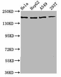
Western Blot. Positive WB detected in: Hela whole cell lysate, HepG2 whole cell lysate, A549 whole cell lysate, 293T whole cell lysate. All lanes: HECW2 antibody at 3.5µg/ml. Secondary. Goat polyclonal to rabbit IgG at 1/50000 dilution. Predicted band size: 176, 136 kDa. Observed band size: 176 kDa.
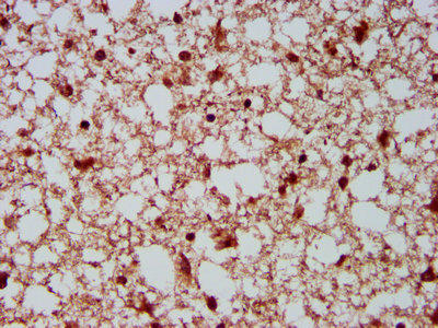
IHC image of PACO59525 diluted at 1:500 and staining in paraffin-embedded human brain tissue performed on a Leica BondTM system. After dewaxing and hydration, antigen retrieval was mediated by high pressure in a citrate buffer (pH 6.0). Section was blocked with 10% normal goat serum 30min at RT. Then primary antibody (1% BSA) was incubated at 4°C overnight. The primary is detected by a biotinylated secondary antibody and visualized using an HRP conjugated SP system.
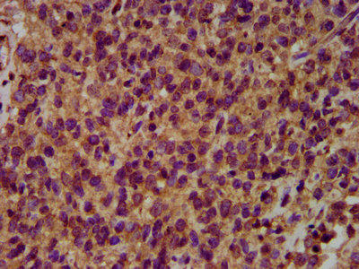
IHC image of PACO59525 diluted at 1:500 and staining in paraffin-embedded human glioma performed on a Leica BondTM system. After dewaxing and hydration, antigen retrieval was mediated by high pressure in a citrate buffer (pH 6.0). Section was blocked with 10% normal goat serum 30min at RT. Then primary antibody (1% BSA) was incubated at 4°C overnight. The primary is detected by a biotinylated secondary antibody and visualized using an HRP conjugated SP system.
Background:
E3 ubiquitin-protein ligase that mediates ubiquitination of TP73. Acts to stabilize TP73 and enhance activation of transcription by TP73. Involved in the regulation of mitotic metaphase/anaphase transition.
HECW2: E3 ubiquitin-protein ligase that mediates ubiquitination of TP73. Acts to stabilize TP73 and enhance activation of transcription by TP73. 2 isoforms of the human protein are produced by alternative splicing.Protein type: Ubiquitin ligase; EC 6.3.2.19; EC 6.3.2.-; Ligase; Ubiquitin conjugating systemChromosomal Location of Human Ortholog: 2q32.3Cellular Component: cytoplasm; nucleusMolecular Function: ubiquitin-protein ligase activityBiological Process: protein ubiquitination during ubiquitin-dependent protein catabolic process


.")
.")

.")

