The c-Fos Antibody (CAB0236) is a high-quality antibody developed for reliable detection and analysis of target proteins. This antibody, produced in rabbits, exhibits high specificity towards human samples and has been validated for use in Western blotting applications. By binding specifically to the FOS protein, this antibody enables precise detection and analysis in a variety of cell types, making it ideal for studies in molecular biology, oncology, and developmental biology.FOS is a crucial regulator of gene expression, playing a central role in cellular processes such as growth, survival, and response to external stimuli.
This antibody is validated for use in WB, IP, ELISA applications and has demonstrated reactivity against Human samples.
Product Name:
c-Fos Antibody
SKU:
CAB0236
Size:
20μL, 100μL
Reactivity:
Human
Conjugate:
Unconjugated
Immunogen:
Recombinant protein (or fragment).This information is considered to be commercially sensitive.
The Fos gene family consists of 4 members: FOS, FOSB, FOSL1, and FOSL2. These genes encode leucine zipper proteins that can dimerize with proteins of the JUN family, thereby forming the transcription factor complex AP-1. As such, the FOS proteins have been implicated as regulators of cell proliferation, differentiation, and transformation. In some cases, expression of the FOS gene has also been associated with apoptotic cell death.
Purification Method
Affinity purification
Gene ID
2353
RRID
AB_2757049
Buffer Information
Store at -20℃. Avoid freeze / thaw cycles. Buffer: PBS containing 50% glycerol, preserved with proclin300 or sodium azide, pH 7.3.
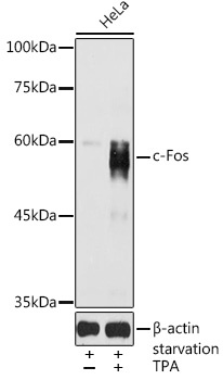
Western blot analysis of lysates from HeLa cells, using c-Fos Rabbit pAb (CAB0236) at 1:1000 dilution. HeLa cells were treated with PMA/TPA (200 nM) at 37℃ for 30 minutes after serum-starvation overnight. Secondary antibody: HRP-conjugated Goat anti-Rabbit IgG (H+L) (CABS014) at 1:10000 dilution. Lysates/proteins: 25μg per lane. Blocking buffer: 3% nonfat dry milk in TBST. Detection: ECL Basic Kit (AbGn00020). Exposure time: 30s.
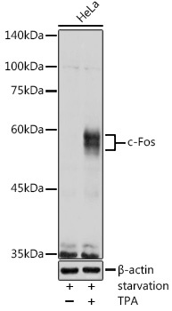
Western blot analysis of lysates from HeLa cells, using c-Fos Rabbit pAb (CAB0236) at 1:1000 dilution. HeLa cells were treated with PMA/TPA (200 nM) at 37℃ for 15 minutes after serum-starvation overnight. Secondary antibody: HRP-conjugated Goat anti-Rabbit IgG (H+L) (CABS014) at 1:10000 dilution. Lysates/proteins: 25μg per lane. Blocking buffer: 3% nonfat dry milk in TBST. Detection: ECL Basic Kit (AbGn00020). Exposure time: 1s.
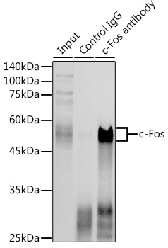
Immunoprecipitation analysis of 300 μg extracts of HeLa cells using 3 μg c-Fos antibody (CAB0236). Western blot was performed from the immunoprecipitate using c-Fos antibody (CAB0236) at a dilution of 1:1000.