The FGFR-Like 1 (FGFRL1) Monoclonal Antibody (PACO56796) is a cutting-edge tool for researchers studying the role of FGFRL1 in various biological processes. FGFRL1 is a member of the fibroblast growth factor receptor (FGFR) family and has been implicated in cell migration, angiogenesis, and development.This monoclonal antibody, produced through rigorous testing and validation processes, specifically targets FGFRL1, allowing for precise detection and analysis in a variety of cell types. It is particularly effective in Western blot applications, providing researchers with reliable data for their experiments.
With FGFRL1's potential involvement in cancer, developmental disorders, and other diseases, this antibody is a valuable asset for scientists conducting research in oncology, developmental biology, and beyond. By understanding the function and regulation of FGFRL1, researchers can uncover new insights into disease pathways and potentially identify targets for therapeutic intervention.
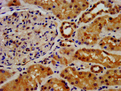
IHC image of PACO56796 diluted at 1:500 and staining in paraffin-embedded human kidney tissue performed on a Leica BondTM system. After dewaxing and hydration, antigen retrieval was mediated by high pressure in a citrate buffer (pH 6.0). Section was blocked with 10% normal goat serum 30min at RT. Then primary antibody (1% BSA) was incubated at 4°C overnight. The primary is detected by a biotinylated secondary antibody and visualized using an HRP conjugated SP system.
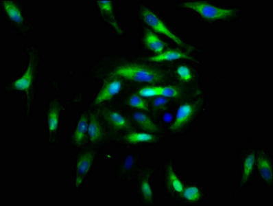
Immunofluorescence staining of Hela cells with PACO56796 at 1:166, counter-stained with DAPI. The cells were fixed in 4% formaldehyde, permeabilized using 0.2% Triton X-100 and blocked in 10% normal Goat Serum. The cells were then incubated with the antibody overnight at 4°C. The secondary antibody was Alexa Fluor 488-congugated AffiniPure Goat Anti-Rabbit IgG(H+L).
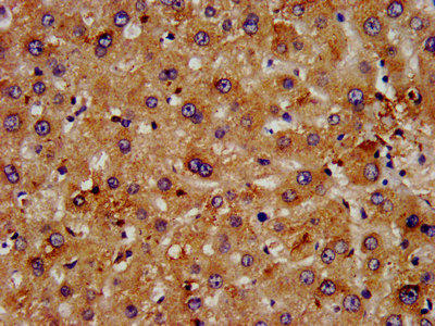
IHC image of PACO56796 diluted at 1:500 and staining in paraffin-embedded human liver tissue performed on a Leica BondTM system. After dewaxing and hydration, antigen retrieval was mediated by high pressure in a citrate buffer (pH 6.0). Section was blocked with 10% normal goat serum 30min at RT. Then primary antibody (1% BSA) was incubated at 4°C overnight. The primary is detected by a biotinylated secondary antibody and visualized using an HRP conjugated SP system.
FGFRL1: Has a negative effect on cell proliferation.Protein type: Membrane protein, integralChromosomal Location of Human Ortholog: 4p16Cellular Component: Golgi apparatus; plasma membrane; transport vesicleMolecular Function: fibroblast growth factor receptor activity; heparin bindingBiological Process: fibroblast growth factor receptor signaling pathway; protein heterooligomerization; protein homooligomerizationDisease: Wolf-hirschhorn Syndrome
UniProt Protein Details:
NCBI Summary:
The protein encoded by this gene is a member of the fibroblast growth factor receptor (FGFR) family, where amino acid sequence is highly conserved between members and throughout evolution. FGFR family members differ from one another in their ligand affinities and tissue distribution. A full-length representative protein would consist of an extracellular region, composed of three immunoglobulin-like domains, a single hydrophobic membrane-spanning segment and a cytoplasmic tyrosine kinase domain. The extracellular portion of the protein interacts with fibroblast growth factors, setting in motion a cascade of downstream signals, ultimately influencing mitogenesis and differentiation. A marked difference between this gene product and the other family members is its lack of a cytoplasmic tyrosine kinase domain. The result is a transmembrane receptor that could interact with other family members and potentially inhibit signaling. Multiple alternatively spliced transcript variants encoding the same isoform have been found for this gene. [provided by RefSeq, Jul 2008]


 (Active) (RPES3508)")
")

