The FATE1 Monoclonal Antibody (PACO38678) is a powerful tool for researchers studying FATE1, a protein involved in cell fate determination and apoptosis. This antibody, generated using innovative technology, specifically recognizes FATE1 and has been validated for use in a variety of experimental techniques, including immunohistochemistry and flow cytometry.FATE1 is a key player in regulating cell survival and death pathways, making it a promising target for investigating cancer progression and therapy resistance.
By using the FATE1 Antibody, researchers can explore the function and expression levels of FATE1 in different cell types and tissues, providing valuable insights into its role in disease pathogenesis.With its high specificity and sensitivity, the FATE1 Monoclonal Antibody offers researchers a reliable tool for advancing their understanding of FATE1 biology and its implications in cancer research and therapeutics. Order now to enhance your research in cell fate determination and apoptosis pathways.
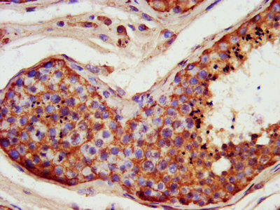
IHC image of PACO38678 diluted at 1:400 and staining in paraffin-embedded human testis tissue performed on a Leica BondTM system. After dewaxing and hydration, antigen retrieval was mediated by high pressure in a citrate buffer (pH 6.0). Section was blocked with 10% normal goat serum 30min at RT. Then primary antibody (1% BSA) was incubated at 4°C overnight. The primary is detected by a biotinylated secondary antibody and visualized using an HRP conjugated SP system.
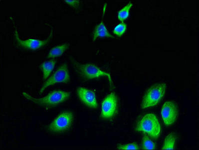
Immunofluorescent analysis of A549 cells using PACO38678 at dilution of 1:100 and Alexa Fluor 488-congugated AffiniPure Goat Anti-Rabbit IgG(H+L).
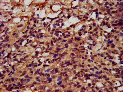
IHC image of PACO38678 diluted at 1:400 and staining in paraffin-embedded human ovarian cancer performed on a Leica BondTM system. After dewaxing and hydration, antigen retrieval was mediated by high pressure in a citrate buffer (pH 6.0). Section was blocked with 10% normal goat serum 30min at RT. Then primary antibody (1% BSA) was incubated at 4°C overnight. The primary is detected by a biotinylated secondary antibody and visualized using an HRP conjugated SP system.
Background:
Involved in the regulation of endoplasmic reticulum (ER)-mitochondria coupling. Negatively regulates the ER-mitochondria distance and Ca2+ transfer from ER to mitochondria possibly implicating it in the regulation of apoptosis. May collaborate with RNF183 to restrain BIK protein levels thus regulating apoptotic signaling.
Synonyms:
Fetal and adult testis-expressed transcript protein (Cancer/testis antigen 43) (CT43) (Tumor antigen BJ-HCC-2), FATE1, FATE
UniProt Protein Function:
FATE1: Protein type: Cancer Testis Antigen (CTA); Membrane protein, integralChromosomal Location of Human Ortholog: Xq28Cellular Component: endoplasmic reticulum; integral to membraneMolecular Function: protein binding
UniProt Protein Details:
NCBI Summary:
This gene encodes a cancer-testis antigen that is highly expressed in hepatocellular carcinomas and other tumors and weakly expressed in normal tissues except testis. The protein is strongly expressed in spermatogonia, primary spermatocytes, and Sertoli cells in seminiferous tubules. This protein may have a role in the control of early testicular differentiation and cell proliferation. [provided by RefSeq, Jan 2010]


.")
.")
. Section was blocked with 10% normal goat serum 30min at RT. Then primary antibody (1% BSA) was incubated at 4°C overnight. The primary is detected by a Goat anti-rabbit IgG labeled by HRP and visualized using 0.05% DAB.")
.")

.")
