The FABP1 Monoclonal Antibody (CAB11213) is a high-quality antibody developed for reliable detection and analysis of target proteins. This antibody, produced using rabbit monoclonal technology, is highly specific and sensitive for detecting FABP1 in human samples. Validated for use in applications such as Western blot, immunohistochemistry, and immunofluorescence, this antibody allows for precise and accurate analysis of FABP1 expression in various tissues and cell types.FABP1, also known as liver-type fatty acid-binding protein, plays a crucial role in intracellular fatty acid transport and metabolism.
This antibody is validated for use in WB, IF/ICC, ELISA, IF-P applications and has demonstrated reactivity against Human, Mouse, Rat samples.
Product Name:
FABP1 Monoclonal Antibody
SKU:
CAB11213
Size:
20μL, 100μL
Reactivity:
Human, Mouse, Rat
Clone Number:
ARC0545
Conjugate:
Unconjugated
Immunogen:
Synthetic peptide. This information is considered to be commercially sensitive.
Recommended starting concentration is 1 μg/mL. Please optimize the concentration based on your specific assay requirements.
Synonyms:
FABPL, L-FABP, FABP1
Positive Sample:
Mouse liver, Mouse small intestine, Rat liver, Hep G2
Cellular Localization:
Cytoplasm.
Calculated MW:
14kDa
Observed MW:
14kDa
This gene encodes the fatty acid binding protein found in liver. Fatty acid binding proteins are a family of small, highly conserved, cytoplasmic proteins that bind long-chain fatty acids and other hydrophobic ligands. This protein and FABP6 (the ileal fatty acid binding protein) are also able to bind bile acids. It is thought that FABPs roles include fatty acid uptake, transport, and metabolism.
Purification Method
Affinity purification
Gene ID
2168
RRID
AB_2861522
Buffer Information
Store at -20℃. Avoid freeze / thaw cycles. Buffer: PBS containing 50% glycerol and 0.05% BSA, preserved with proclin300 or sodium azide, pH 7.3.
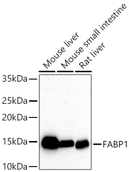
Western blot analysis of various lysates using FABP1 Rabbit mAb (CAB11213) at 1:1000 dilution incubated at room temperature for 1.5 hours. Secondary antibody: HRP-conjugated Goat anti-Rabbit IgG (H+L) (CABS014) at 1:10000 dilution. Lysates/proteins: 25 μg per lane. Blocking buffer: 3% nonfat dry milk in TBST. Detection: ECL Basic Kit (AbGn00020). Exposure time: 30s.
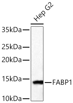
Western blot analysis of lysates from Hep G2 cells using FABP1 Rabbit mAb (CAB11213) at 1:1000 dilution incubated at room temperature for 1.5 hours. Secondary antibody: HRP-conjugated Goat anti-Rabbit IgG (H+L) (CABS014) at 1:10000 dilution. Lysates/proteins: 25 μg per lane. Blocking buffer: 3% nonfat dry milk in TBST. Detection: ECL Basic Kit (AbGn00020). Exposure time: 90s.
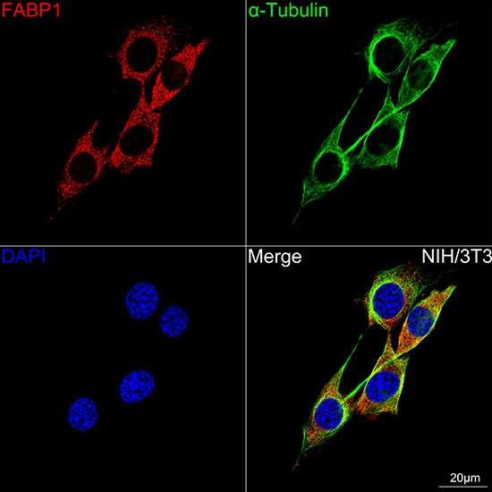
Confocal imaging of NIH/3T3 cells using FABP1 Rabbit mAb (CAB11213, dilution 1:100) followed by a further incubation with Cy3 Goat Anti-Rabbit IgG (H+L) (CABS007, dilution 1:500) (Red). The cells were counterstained with α-Tubulin Mouse mAb (AC012, dilution 1:400) followed by incubation with ABflo® 488-conjugated Goat Anti-Mouse IgG (H+L) Ab (CABS076, dilution 1:500) (Green). DAPI was used for nuclear staining (Blue). Objective: 100x.
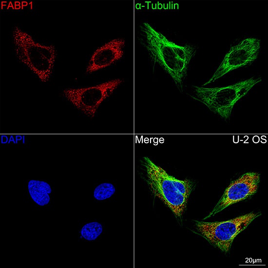
Confocal imaging of U-2 OS cells using FABP1 Rabbit mAb (CAB11213, dilution 1:100) followed by a further incubation with Cy3 Goat Anti-Rabbit IgG (H+L) (CABS007, dilution 1:500) (Red). The cells were counterstained with α-Tubulin Mouse mAb (AC012, dilution 1:400) followed by incubation with ABflo® 488-conjugated Goat Anti-Mouse IgG (H+L) Ab (CABS076, dilution 1:500) (Green). DAPI was used for nuclear staining (Blue). Objective: 100x.


![Anti-FABP1 [8G6-D5] Monoclonal Antibody, capture (AGMB05449)](https://cdn11.bigcommerce.com/s-h68l9z2lnx/images/stencil/590x590/products/276733/677551/anti-fabp1-8g6-d5-monoclonal-antibody-capture-agmb05449__51283.1773032920.jpg?c=2 "Anti-FABP1 [8G6-D5] Monoclonal Antibody, capture (AGMB05449)")
![Anti-FABP1 (2D6) [2D6-F6] Monoclonal Antibody (AGMB04492)](https://cdn11.bigcommerce.com/s-h68l9z2lnx/images/stencil/590x590/products/275780/676259/anti-fabp1-2d6-2d6-f6-monoclonal-antibody-agmb04492__62738.1773028839.jpg?c=2 "Anti-FABP1 (2D6) [2D6-F6] Monoclonal Antibody (AGMB04492)")
![Anti-FABP1 (8G6) [8G6-D5] Monoclonal Antibody (AGMB04493)](https://cdn11.bigcommerce.com/s-h68l9z2lnx/images/stencil/590x590/products/275781/679849/anti-fabp1-8g6-8g6-d5-monoclonal-antibody-agmb04493__19125.1773040224.jpg?c=2 "Anti-FABP1 (8G6) [8G6-D5] Monoclonal Antibody (AGMB04493)")
![Anti-FABP1 [2D6-F6] Monoclonal Antibody, unconjugated, detector (AGMB05450)](https://cdn11.bigcommerce.com/s-h68l9z2lnx/images/stencil/590x590/products/276734/679813/anti-fabp1-2d6-f6-monoclonal-antibody-unconjugated-detector-agmb05450__44079.1773040107.jpg?c=2 "Anti-FABP1 [2D6-F6] Monoclonal Antibody, unconjugated, detector (AGMB05450)")