Estrogen Receptor-alpha (Phospho-Ser106) Colorimetric Cell-Based ELISA Kit
- SKU:
- CBCAB01525
- Product Type:
- ELISA Kit
- ELISA Type:
- Cell Based Phospho Specific
- Research Area:
- Epigenetics and Nuclear Signaling
- Reactivity:
- Human
- Mouse
- Rat
- Detection Method:
- Colorimetric
Description
Estrogen Receptor-alpha (Phospho-Ser106)Colorimetric Cell-Based ELISA Kit
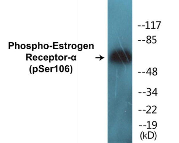
The Estrogen Receptor Alpha Phospho-Ser106 Colorimetric Cell-Based ELISA Kit is a cutting-edge tool for researchers looking to study the phosphorylation status of estrogen receptor alpha at serine 106 in cell-based assays. This kit offers high sensitivity and specificity, providing accurate and reliable results for detecting changes in ERα phosphorylation levels.Estrogen receptor alpha is a key player in mediating the effects of estrogen on gene expression and cell growth, and its phosphorylation at specific sites can regulate its activity. Phosphorylation of ERα at serine 106 has been linked to various physiological and pathological processes, including breast cancer and other hormone-related disorders.
By using this ELISA kit, researchers can gain valuable insights into the role of ERα phosphorylation in normal physiological processes and disease states, paving the way for the development of novel therapeutic strategies targeting estrogen signaling pathways. With its user-friendly protocol and robust performance, this kit is a valuable tool for advancing research in the field of hormone signaling and estrogen-related diseases.
| Product Name: | Estrogen Receptor-alpha (Phospho-Ser106) Colorimetric Cell-Based ELISA |
| Product Code: | CBCAB01525 |
| ELISA Type: | Cell-Based |
| Target: | Estrogen Receptor-alpha (Phospho-Ser106) |
| Reactivity: | Human, Mouse, Rat |
| Dynamic Range: | > 5000 Cells |
| Detection Method: | Colorimetric 450 nm |
| Format: | 2 x 96-Well Microplates |
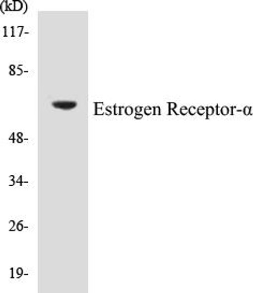
The Estrogen Receptor-alpha (Phospho-Ser106) Colorimetric Cell-Based ELISA Kit is a convenient, lysate-free, high throughput and sensitive assay kit that can detect Estrogen Receptor-alpha protein phosphorylation and expression profile in cells. The kit can be used for measuring the relative amounts of phosphorylated Estrogen Receptor-alpha in cultured cells as well as screening for the effects that various treatments, inhibitors (ie. siRNA or chemicals), or activators have on Estrogen Receptor-alpha phosphorylation.
Qualitative determination of Estrogen Receptor-alpha (Phospho-Ser106) concentration is achieved by an indirect ELISA format. In essence, Estrogen Receptor-alpha (Phospho-Ser106) is captured by Estrogen Receptor-alpha (Phospho-Ser106)-specific primary (1ø) antibodies while the HRP-conjugated secondary (2ø) antibodies bind the Fc region of the 1ø antibody. Through this binding, the HRP enzyme conjugated to the 2ø antibody can catalyze a colorimetric reaction upon substrate addition. Due to the qualitative nature of the Cell-Based ELISA, multiple normalization methods are needed:
| 1. | A monoclonal antibody specific for human GAPDH is included to serve as an internal positive control in normalizing the target absorbance values. |
| 2. | Following the colorimetric measurement of HRP activity via substrate addition, the Crystal Violet whole-cell staining method may be used to determine cell density. After staining, the results can be analysed by normalizing the absorbance values to cell amounts, by which the plating difference can be adjusted. |
| Database Information: | Gene ID: 2099, UniProt ID: P03372, OMIM: 133430, Unigene: Hs.208124 |
| Gene Symbol: | ESR1 |
| Sub Type: | Phospho |
| UniProt Protein Function: | Function: Nuclear hormone receptor. The steroid hormones and their receptors are involved in the regulation of eukaryotic gene expression and affect cellular proliferation and differentiation in target tissues. Ligand-dependent nuclear transactivation involves either direct homodimer binding to a palindromic estrogen response element (ERE) sequence or association with other DNA-binding transcription factors, such as AP-1/c-Jun, c-Fos, ATF-2, Sp1 and Sp3, to mediate ERE-independent signaling. Ligand binding induces a conformational change allowing subsequent or combinatorial association with multiprotein coactivator complexes through LXXLL motifs of their respective components. Mutual transrepression occurs between the estrogen receptor (ER) and NF-kappa-B in a cell-type specific manner. Decreases NF-kappa-B DNA-binding activity and inhibits NF-kappa-B-mediated transcription from the IL6 promoter and displace RELA/p65 and associated coregulators from the promoter. Recruited to the NF-kappa-B response element of the CCL2 and IL8 promoters and can displace CREBBP. Present with NF-kappa-B components RELA/p65 and NFKB1/p50 on ERE sequences. Can also act synergistically with NF-kappa-B to activate transcription involving respective recruitment adjacent response elements; the function involves CREBBP. Can activate the transcriptional activity of TFF1. Also mediates membrane-initiated estrogen signaling involving various kinase cascades. Isoform 3 is involved in activation of NOS3 and endothelial nitric oxide production. Isoforms lacking one or several functional domains are thought to modulate transcriptional activity by competitive ligand or DNA binding and/or heterodimerization with the full length receptor. Isoform 3 can bind to ERE and inhibit isoform 1. Ref.17 Ref.19 Ref.24 Ref.30 Ref.31 Ref.33 Ref.36 Ref.44 Ref.46 Ref.47 Ref.48 Ref.52 Ref.63 Ref.65 Ref.70 Ref.75 Ref.77 Ref.78 Ref.80 |
| UniProt Protein Details: | Subunit structure: Binds DNA as a homodimer. Can form a heterodimer with ESR2. Isoform 3 can probably homodimerize or heterodimerize with isoform 1 and ESR2. Interacts with FOXC2, MAP1S, SLC30A9, UBE1C and NCOA3 coactivator By similarity. Interacts with EP300; the interaction is estrogen-dependent and enhanced by CITED1. Interacts with CITED1; the interaction is estrogen-dependent. Interacts with NCOA5 and NCOA6 coactivators. Interacts with NCOA7; the interaction is a ligand-inducible. Interacts with PHB2, PELP1 and UBE1C. Interacts with AKAP13. Interacts with CUEDC2. Interacts with KDM5A. Interacts with SMARD1. Interacts with HEXIM1. Interacts with PBXIP1. Interaction with MUC1 is stimulated by 7 beta-estradiol (E2) and enhances ERS1-mediated transcription. Interacts with DNTTIP2, FAM120B and UIMC1. Interacts with isoform 4 of TXNRD1. Interacts with KMT2D/MLL2. Interacts with ATAD2 and this interaction is enhanced by estradiol. Interacts with KIF18A and LDB1. Interacts with RLIM (via C-terminus). Interacts with MACROD1. Interacts with SH2D4A and PLCG. Interaction with SH2D4A blocks binding to PLCG and inhibits estrogen-induced cell proliferation. Interacts with DYNLL1. Interacts with CCDC62 in the presence of estradiol/E2; this interaction seems to enhance the transcription of target genes. Interacts with NR2C1; the interaction prevents homodimerization of ESR1 and suppresses its transcriptional activity and cell growth. Interacts with DYX1C1. Interacts with PRMT2. Interacts with PI3KR1 or PI3KR2, SRC and PTK2/FAK1. Interacts with RBFOX2. Interacts with STK3/MST2 only in the presence of SAV1 and vice-versa. Binds to CSNK1D. Interacts with NCOA2; NCOA2 can interact with ESE1 AF-1 and AF-2 domains simultaneously and mediate their transcriptional synergy. Interacts with DDX5. Interacts with NCOA1; the interaction seems to require a self-association of N-terminal and C-terminal regions. Interacts with ZNF366, DDX17, NFKB1, RELA, SP1 and SP3. Interacts with NRIP1 By similarity. Interacts with GPER1; the interaction occurs in an estrogen-dependent manner. Ref.18 Ref.19 Ref.21 Ref.24 Ref.25 Ref.27 Ref.28 Ref.29 Ref.30 Ref.31 Ref.32 Ref.33 Ref.34 Ref.35 Ref.36 Ref.37 Ref.38 Ref.39 Ref.40 Ref.41 Ref.42 Ref.43 Ref.45 Ref.48 Ref.50 Ref.51 Ref.53 Ref.54 Ref.55 Ref.56 Ref.57 Ref.58 Ref.59 Ref.60 Ref.61 Ref.62 Ref.64 Ref.65 Ref.66 Ref.67 Ref.68 Ref.69 Ref.70 Ref.71 Ref.72 Ref.73 Ref.74 Ref.76 Ref.91 Subcellular location: Isoform 1: Nucleus. Cytoplasm. Cell membrane; Peripheral membrane protein; Cytoplasmic side. Note: A minor fraction is associated with the inner membrane. Ref.6 Ref.22 Ref.48 Ref.59 Ref.64 Ref.77 Ref.79Isoform 3: Nucleus. Cytoplasm. Cell membrane; Peripheral membrane protein; Cytoplasmic side. Cell membrane; Single-pass type I membrane protein. Note: Associated with the inner membrane via palmitoylation Probable. At least a subset exists as a transmembrane protein with a N-terminal extracellular domain. Ref.6 Ref.22 Ref.48 Ref.59 Ref.64 Ref.77 Ref.79Nucleus. Golgi apparatus. Cell membrane. Note: Colocalizes with ZDHHC7 and ZDHHC21 in the Golgi apparatus where most probably palmitoylation occurs. Associated with the plasma membrane when palmitoylated. Ref.6 Ref.22 Ref.48 Ref.59 Ref.64 Ref.77 Ref.79 Tissue specificity: Widely expressed. Isoform 3 is not expressed in the pituitary gland. Ref.19 Domain: Composed of three domains: a modulating N-terminal domain, a DNA-binding domain and a C-terminal ligand-binding domain. The modulating domain, also known as A/B or AF-1 domain has a ligand-independent transactivation function. The C-terminus contains a ligand-dependent transactivation domain, also known as E/F or AF-2 domain which overlaps with the ligand binding domain. AF-1 and AF-2 activate transcription independently and synergistically and act in a promoter- and cell-specific manner. AF-1 seems to provide the major transactivation function in differentiated cells. Post-translational modification: Phosphorylated by cyclin A/CDK2 and CK1. Phosphorylation probably enhances transcriptional activity. Self-association induces phosphorylation. Dephosphorylation at Ser-118 by PPP5C inhibits its transactivation activity. Ref.10 Ref.14 Ref.16 Ref.26 Ref.46 Ref.72Glycosylated; contains N-acetylglucosamine, probably O-linked. Ref.23Ubiquitinated. Deubiquitinated by OTUB1. Ref.71Dimethylated by PRMT1 at Arg-260. The methylation may favor cytoplasmic localization. Ref.64Palmitoylated (isoform 3) Not biotinylated (isoform 3) Ref.22 Ref.79Palmitoylated by ZDHHC7 and ZDHHC21. Palmitoylation is required for plasma membrane targeting and for rapid intracellular signaling via ERK and AKT kinases and cAMP generation, but not for signaling mediated by the nuclear hormone receptor. Ref.22 Ref.79 Polymorphism: Genetic variations in ESR1 are correlated with bone mineral density (BMD). Low BMD is a risk factor for osteoporotic fracture. Osteoporosis is characterized by reduced bone mineral density, disrutption of bone microarchitecture, and the alteration of the amount and variety of non-collagenous proteins in bone. Osteoporotic bones are more at risk of fracture. Involvement in Disease: Estrogen resistance (ESTRR) [MIM:615363]: A disorder characterized by partial or complete resistance to estrogens, in the presence of elevated estrogen serum levels. Clinical features include absence of the pubertal growth spurt, delayed bone maturation, unfused epiphyses, reduced bone mineral density, osteoporosis, continued growth into adulthood and very tall adult stature. Glucose intolerance, hyperinsulinemia and lipid abnormalities may also be present.Note: The disease is caused by mutations affecting the gene represented in this entry. Ref.88 Ref.93 Miscellaneous: Selective estrogen receptor modulators (SERMs), such as tamoxifen, raloxifene, toremifene, lasofoxifene, clomifene, femarelle and ormeloxifene, have tissue selective agonistic and antagonistic effects on the estrogen receptor (ER). They interfere with the ER association with coactivators or corepressors, mainly involving the AF-2 domain. Sequence similarities: Belongs to the nuclear hormone receptor family. NR3 subfamily.Contains 1 nuclear receptor DNA-binding domain. |
| NCBI Summary: | This gene encodes an estrogen receptor, a ligand-activated transcription factor composed of several domains important for hormone binding, DNA binding, and activation of transcription. The protein localizes to the nucleus where it may form a homodimer or a heterodimer with estrogen receptor 2. Estrogen and its receptors are essential for sexual development and reproductive function, but also play a role in other tissues such as bone. Estrogen receptors are also involved in pathological processes including breast cancer, endometrial cancer, and osteoporosis. Alternative splicing results in several transcript variants, which differ in their 5' UTRs and use different promoters. [provided by RefSeq, Jul 2008] |
| UniProt Code: | P03372 |
| NCBI GenInfo Identifier: | 544257 |
| NCBI Gene ID: | 2099 |
| NCBI Accession: | P03372.2 |
| UniProt Secondary Accession: | P03372,Q13511, Q14276, Q5T5H7, Q6MZQ9, Q9NU51, Q9UDZ7 Q9UIS7, |
| UniProt Related Accession: | P03372 |
| Molecular Weight: | |
| NCBI Full Name: | Estrogen receptor |
| NCBI Synonym Full Names: | estrogen receptor 1 |
| NCBI Official Symbol: | ESR1 |
| NCBI Official Synonym Symbols: | ER; ESR; Era; ESRA; ESTRR; NR3A1 |
| NCBI Protein Information: | estrogen receptor; ER-alpha; estradiol receptor; estrogen nuclear receptor alpha; estrogen receptor alpha E1-E2-1-2; estrogen receptor alpha E1-N2-E2-1-2; estrogen receptor alpha delta 4 +49 isoform; nuclear receptor subfamily 3 group A member 1; estrogen receptor alpha 3*,4,5,6,7*/822 isoform; estrogen receptor alpha delta 4*,5,6,7*/654 isoform; estrogen receptor alpha delta 4*,5,6,7,8*/901 isoform; estrogen receptor alpha delta 3*,4,5,6,7*/819-2 isoform; estrogen receptor alpha delta 3*,4,5,6,7*,8*/941 isoform |
| UniProt Protein Name: | Estrogen receptor |
| UniProt Synonym Protein Names: | ER-alpha; Estradiol receptor; Nuclear receptor subfamily 3 group A member 1 |
| Protein Family: | Erbin |
| UniProt Gene Name: | ESR1 |
| UniProt Entry Name: | ESR1_HUMAN |
| Component | Quantity |
| 96-Well Cell Culture Clear-Bottom Microplate | 2 plates |
| 10X TBS | 24 mL |
| Quenching Buffer | 24 mL |
| Blocking Buffer | 50 mL |
| 15X Wash Buffer | 50 mL |
| Primary Antibody Diluent | 12 mL |
| 100x Anti-Phospho Target Antibody | 60 µL |
| 100x Anti-Target Antibody | 60 µL |
| Anti-GAPDH Antibody | 60 µL |
| HRP-Conjugated Anti-Rabbit IgG Antibody | 12 mL |
| HRP-Conjugated Anti-Mouse IgG Antibody | 12 mL |
| SDS Solution | 12 mL |
| Stop Solution | 24 mL |
| Ready-to-Use Substrate | 12 mL |
| Crystal Violet Solution | 12 mL |
| Adhesive Plate Seals | 2 seals |
The following materials and/or equipment are NOT provided in this kit but are necessary to successfully conduct the experiment:
- Microplate reader able to measure absorbance at 450 nm and/or 595 nm for Crystal Violet Cell Staining (Optional)
- Micropipettes with capability of measuring volumes ranging from 1 µL to 1 ml
- 37% formaldehyde (Sigma Cat# F-8775) or formaldehyde from other sources
- Squirt bottle, manifold dispenser, multichannel pipette reservoir or automated microplate washer
- Graph paper or computer software capable of generating or displaying logarithmic functions
- Absorbent papers or vacuum aspirator
- Test tubes or microfuge tubes capable of storing ≥1 ml
- Poly-L-Lysine (Sigma Cat# P4832 for suspension cells)
- Orbital shaker (optional)
- Deionized or sterile water
*Note: Protocols are specific to each batch/lot. For the correct instructions please follow the protocol included in your kit.
| Step | Procedure |
| 1. | Seed 200 µL of 20,000 adherent cells in culture medium in each well of a 96-well plate. The plates included in the kit are sterile and treated for cell culture. For suspension cells and loosely attached cells, coat the plates with 100 µL of 10 µg/ml Poly-L-Lysine (not included) to each well of a 96-well plate for 30 minutes at 37 °C prior to adding cells. |
| 2. | Incubate the cells for overnight at 37 °C, 5% CO2. |
| 3. | Treat the cells as desired. |
| 4. | Remove the cell culture medium and rinse with 200 µL of 1x TBS, twice. |
| 5. | Fix the cells by incubating with 100 µL of Fixing Solution for 20 minutes at room temperature. The 4% formaldehyde is used for adherent cells and 8% formaldehyde is used for suspension cells and loosely attached cells. |
| 6. | Remove the Fixing Solution and wash the plate 3 times with 200 µL 1x Wash Buffer for five minutes each time with gentle shaking on the orbital shaker. The plate can be stored at 4 °C for a week. |
| 7. | Add 100 µL of Quenching Buffer and incubate for 20 minutes at room temperature. |
| 8. | Wash the plate 3 times with 1x Wash Buffer for 5 minutes each time. |
| 9. | Add 200 µL of Blocking Buffer and incubate for 1 hour at room temperature. |
| 10. | Wash 3 times with 200 µL of 1x Wash Buffer for 5 minutes each time. |
| 11. | Add 50 µL of 1x primary antibodies Anti-Estrogen Receptor-alpha (Phospho-Ser106) Antibody, Anti-Estrogen Receptor-alpha Antibody and/or Anti-GAPDH Antibody) to the corresponding wells, cover with Parafilm and incubate for 16 hours (overnight) at 4 °C. If the target expression is known to be high, incubate for 2 hours at room temperature. |
| 12. | Wash 3 times with 200 µL of 1x Wash Buffer for 5 minutes each time. |
| 13. | Add 50 µL of 1x secondary antibodies (HRP-Conjugated AntiRabbit IgG Antibody or HRP-Conjugated Anti-Mouse IgG Antibody) to corresponding wells and incubate for 1.5 hours at room temperature. |
| 14. | Wash 3 times with 200 µL of 1x Wash Buffer for 5 minutes each time. |
| 15. | Add 50 µL of Ready-to-Use Substrate to each well and incubate for 30 minutes at room temperature in the dark. |
| 16. | Add 50 µL of Stop Solution to each well and read OD at 450 nm immediately using the microplate reader. |
(Additional Crystal Violet staining may be performed if desired – details of this may be found in the kit technical manual.)


