Description
ERK Phosphorylation Assay
The mitogen-activated protein kinase (MAPK/ERK) pathway plays a key role in cell proliferation, differentiation and migration. Stimulation by mitogens eventually leads to phosphorylation of ERK1 (T202/Y204) and ERK2 (T185/Y187). The MAPK/ERK cascade presents many interesting drug targets for the development of cancer therapies.
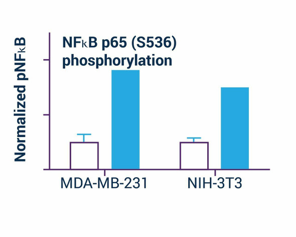
Assay Genie's cell-based ELISA ERK Phosphorylation Assay measures dually phosphorylated ERK1/2 in whole cells and normalizes the signal to the total protein content. This simple and efficient assay eliminates the need for cell lysate preparation and can be used to study kinase signaling and the effects of kinase inhibitors on cells. In this assay, cells are grown in 96-well plates and treated with ligands or drugs. Cells are then fixed and permeabilized in the wells. ERK1/2 phosphorylation (pERK) using a fluorescent ELISA followed by total protein measurement in each well.Applications
For quantitative fluorescent immunoenzymatic assay of ERK1/2 phosphorylation status in cultured cells.Key Features
- New and improved. Total assay time reduced from the standard 21 hours to 6.5 hours (hands-on time 2.5 hrs).
- Simple and convenient. Cells are directly cultured in 96-well plates. No cell lysis necessary.
- Accurate and high-throughput. Protein phosphorylation is normalized to total cellular protein in the same well, greatly minimizing well-to-well variations. Can be readily automated as a high-throughput 96-well plate assay for thousands of samples per day.
ERK Phosphorylation Assay Information:
| Kit Includes: | 10x Wash Buffer: 25 mL Blocking Buffer: 25 mL Protein Stain: 6 mL HRP Substrate: 6 mL HRP-Ab2: 10 uL pERK-Ab1: 10 uL |
| Kit Requires: | 37% formaldehyde; 3% H2O2; black cell culture 96-well plate; plate sealers; deionized or distilled water; pipetting devices; cell culture incubators; centrifuge tubes; fluorescence plate reader capable of reading at ex/em = 530/585 nm and at ex/em =360/450 nm. |
| Method of Detection: | FL360/450nm, 530/585nm |
| Samples: | Cell, tissue etc |
| Species: | Human, mouse, rat. |
| Protocol Length: | Assay takes 6.5 hrs, hands-on time 2.5 hrs |
| Size: | 100 tests |
| Storage: | Store all reagents at -20°C |
| Shelf Life: | 6 months |


