The CDH7 Antibody (PAC057300) is a valuable tool for researchers studying the CDH7 protein, a cell adhesion molecule implicated in various biological processes. This polyclonal antibody, produced in rabbits, exhibits high reactivity with human samples and is ideal for use in Western blot applications. By binding specifically to CDH7, this antibody enables the detection and analysis of CDH7 protein in a variety of cell types.CDH7, also known as cadherin-7, plays a crucial role in cell-cell adhesion and tissue organization, making it a key player in development and disease processes.
Its involvement in cell signaling and migration also suggests potential implications in cancer metastasis and tissue remodeling. Studying the function of CDH7 can provide valuable insights into these processes and may offer new therapeutic strategies for cancer and other diseases. Overall, the CDH7 Antibody offers researchers a reliable tool for investigating the role of CDH7 in various biological contexts and advancing our understanding of its potential implications in health and disease.
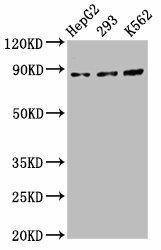
Western Blot. Positive WB detected in: HepG2 whole cell lysate, 293 whole cell lysate, K562 whole cell lysate. All lanes: CDH7 antibody at 5.5µg/ml. Secondary. Goat polyclonal to rabbit IgG at 1/50000 dilution. Predicted band size: 88 kDa. Observed band size: 88 kDa.
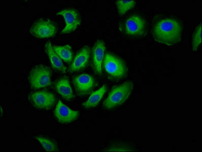
Immunofluorescence staining of A549 cells with PACO57300 at 1:133, counter-stained with DAPI. The cells were fixed in 4% formaldehyde, permeabilized using 0.2% Triton X-100 and blocked in 10% normal Goat Serum. The cells were then incubated with the antibody overnight at 4°C. The secondary antibody was Alexa Fluor 488-congugated AffiniPure Goat Anti-Rabbit IgG(H+L).
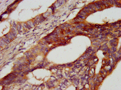
IHC image of PACO57300 diluted at 1:400 and staining in paraffin-embedded human colon cancer performed on a Leica BondTM system. After dewaxing and hydration, antigen retrieval was mediated by high pressure in a citrate buffer (pH 6.0). Section was blocked with 10% normal goat serum 30min at RT. Then primary antibody (1% BSA) was incubated at 4°C overnight. The primary is detected by a biotinylated secondary antibody and visualized using an HRP conjugated SP system.
Background:
Cadherins are calcium-dependent cell adhesion proteins. They preferentially interact with themselves in a homophilic manner in connecting cells; cadherins may thus contribute to the sorting of heterogeneous cell types.
Synonyms:
Cadherin-7, CDH7, CDH7L1
UniProt Protein Function:
CDH7: Cadherins are calcium-dependent cell adhesion proteins. They preferentially interact with themselves in a homophilic manner in connecting cells; cadherins may thus contribute to the sorting of heterogeneous cell types.
UniProt Protein Details:
Protein type:Cell adhesion; Membrane protein, integral
Chromosomal Location of Human Ortholog: 18q22.1
Cellular Component: plasma membrane
NCBI Summary:
This gene encodes a type II classical cadherin of the cadherin superfamily. Alternative splicing results in multiple transcript variants, at least one of which encodes a preproprotein that is proteolytically processed to generate the mature glycoprotein. This calcium dependent cell-cell adhesion molecule is comprised of five extracellular cadherin repeats, a transmembrane region and a highly conserved cytoplasmic tail. Type II (atypical) cadherins are defined based on their lack of a histidine-alanine-valine (HAV) cell adhesion recognition sequence specific to type I cadherins. Cadherins mediate cell-cell binding in a homophilic manner, contributing to the sorting of heterogeneous cell types. Mutations in this gene may be associated with bipolar disease in human patients. This gene is present in a gene cluster on chromosome 18. [provided by RefSeq, May 2016]


 ELISA Kit (MOEB1084)")
 ELISA Kit (HUEB1320)")

.")