The Functions of Microglial Cells & Their Role in Neurodegenerative Disorders
Key Takeaways:
- Microglia are immune cells in the CNS, originating from myeloid precursor cells.
- They surveil the CNS, respond to changes, and maintain neural health.
- Microglia exist in different states: amoeboid, ramified, and reactive.
- Their activation is crucial in neurodegenerative diseases and brain health.
What are Microglia?
Microglia are a specialized type of immune cells that reside within the central nervous system (CNS), including the brain and spinal cord. Originating from myeloid precursor cells, microglia colonize the CNS during early development. In their resting state, microglia exhibit a distinctive morphology with small cell bodies and highly branched processes. Strategically positioned throughout the brain, they continually survey the neuronal environment. Equipped with an array of receptors, microglia can detect changes and abnormalities in their surroundings. Microglia can rapidly transition from a quiescent state to an activated state, undergoing morphological and functional transformations.
Schematic of Microglia
What is the Function of Microglia?
Microglia serve a myriad of functions within the central nervous system, contributing to the overall health and functioning of neural tissue. Microglia constantly surveil the CNS for signs of injury, infection, or any aberrant changes. Their ability to detect and respond to various signals, including neurotransmitters and cytokines, allows them to auickly respond to disturbances. Additionally, microglia play a crucial role in the clearance of cellular debris, damaged neurons, and toxic protein aggregates through phagocytosis. By efficiently removing these harmful substances, microglia help maintain a clean and healthy neuronal environment.
Furthermore, microglia actively participate in synaptic pruning during brain development and remodeling in adulthood, ensuring optimal connectivity and functioning of neural circuits. They also possess immunomodulatory properties, releasing cytokines, chemokines, and growth factors that influence the activity of other immune cells and facilitate tissue repair. However, dysregulated or chronic activation of microglia can contribute to neuroinflammation, leading to neuronal damage and neurodegenerative disorders.
What Are the Types of Microglia?
Microglial cells exhibit different morphological and functional states, contributing to their diverse roles in the central nervous system. One classification scheme recognizes several distinct types of microglia. There are three main types of microglial cells: amoeboid microglia, ramified microglia, and reactive microglia. The remarkable plasticity of microglial cells allows them to adapt and respond to their microenvironment, contributing to immune surveillance, neuroinflammation, tissue repair, and synaptic remodeling.
Amoeboid Microglia:
Amoeboid microglia represent an activated state characterized by a rounded morphology with retracting processes. They are highly mobile and phagocytic, playing a crucial role in clearing cellular debris and promoting tissue repair. Amoeboid microglia are typically observed during early development or in response to injury or inflammation. These dynamic cells swiftly respond to changes in their environment, transitioning from a resting state to an activated state. Their amoeboid shape allows them to efficiently migrate through the tissue and engulf debris, contributing to the resolution of injury or inflammation. Amoeboid microglia release various signaling molecules and pro-inflammatory cytokines, participating in immune responses within the central nervous system.
Ramified Microglia:
Ramified microglia, also known as resting microglia, are the baseline state of microglia in the healthy central nervous system. They have a highly branched morphology, with numerous processes extending from their cell bodies. Ramified microglia continuously monitor the neuronal environment, ensuring the maintenance of homeostasis. In this resting state, they play a vital role in immune surveillance, actively surveying the surrounding neural tissue for any abnormalities. Ramified microglia constantly interact with neurons and other glial cells, contributing to neuronal health and supporting the normal functioning of the brain. Although they are considered resting, they retain the ability to respond rapidly to any perturbations by transitioning into an activated state.
Reactive Microglia:
Reactive microglia encompass various activated states of microglia, displaying diverse phenotypes and functional profiles. These cells are involved in immune responses and can exhibit different morphologies beyond the typical amoeboid form. Reactive microglia release both pro-inflammatory and anti-inflammatory cytokines, participate in phagocytosis, and modulate the immune response within the central nervous system. Their activation is often observed in the context of neurodegenerative disorders, where they become involved in the clearance of toxic protein aggregates and the promotion of tissue repair. The functional profile of reactive microglia can vary depending on the specific stimulus or neurodegenerative context, highlighting their dynamic nature and adaptability.
What Activates Microglia?
Activation of microglia occurs in response to various stimuli, including pathogen-associated molecular patterns (PAMPs) and damage-associated molecular patterns (DAMPs). Microglial activation involves a complex interplay between PAMPs, DAMPs, and their corresponding receptors, triggering intracellular signaling pathways and gene expression changes. The activation process can result in either a pro-inflammatory (M1) phenotype or an anti-inflammatory and tissue repair (M2) phenotype, depending on the context and microenvironment.
Markers of Microglial Activation
Microglial activation plays a crucial role in the immune response within the CNS and is associated with various neurodegenerative disorders. Several markers are commonly used to identify and characterize activated microglia. There are a number of markers that can be used to detect activated microglia. These markers include CD11b, CD68, Iba-. Activated microglia can be detected by using imaging techniques such as positron emission tomography (PET), magnetic resonance imaging (MRI), and single-photon emission computed tomography (SPECT).
-
CD68: CD68 is a glycoprotein predominantly expressed by activated microglia and macrophages. It is commonly used as a marker for phagocytic activity, indicating the presence of activated microglia involved in clearing cellular debris and pathogens. CD68 expression increases in response to injury, infection, or neurodegenerative processes, reflecting the role of microglia in immune surveillance and phagocytosis.
-
Ionized calcium-binding adapter molecule 1 (Iba1): Iba1 is a calcium-binding protein highly expressed in microglia. It is often used as a general marker of microglial activation. Iba1 is involved in microglial motility, phagocytosis, and cytoskeletal rearrangements. Increased expression of Iba1 indicates microglial activation and their transition into an amoeboid or reactive state.
-
CD11b: CD11b is a cell surface antigen expressed by microglia and other myeloid cells. It plays a role in cell adhesion and phagocytosis. Upregulation of CD11b is associated with microglial activation and their involvement in neuroinflammatory processes. CD11b is frequently used as a marker to identify activated microglia in experimental studies.
-
Major Histocompatibility Complex II (MHC-II): MHC-II molecules are involved in antigen presentation and immune response modulation. Upregulation of MHC-II on microglia is indicative of their activation and involvement in immune surveillance and antigen presentation to T-cells. Increased expression of MHC-II is often observed in neuroinflammatory conditions and neurodegenerative disorders.
-
C-X3-C chemokine receptor 1 (CX3CR1): CX3CR1 is a chemokine receptor predominantly expressed on microglia. It plays a role in microglial migration, cell-to-cell communication, and neuronal interactions. Downregulation of CX3CR1 has been associated with microglial activation and neuroinflammatory processes in various diseases.
Micoglia and Brain Health
One crucial aspect of microglial function is their proficiency in phagocytosis and clearance. Microglia are responsible for engulfing and removing cellular debris, apoptotic cells, and toxic protein aggregates from the brain. This phagocytic activity is essential for maintaining a healthy neuronal environment and preventing the accumulation of potentially harmful substances. By efficiently clearing cellular waste, microglia contribute to the overall well-being and functionality of the CNS.
Microglia actively participate in the refinement and remodeling of synaptic connections during brain development and throughout life. They interact with synapses, regulating synaptic pruning and plasticity. Microglia's involvement in synaptic maintenance ensures the optimal functioning of neuronal circuits, promoting learning, memory formation, and neuronal connectivity.
Another critical role of microglia is their contribution to the regulation of immune responses and inflammation within the CNS. They can transition into an activated state in response to various stimuli, releasing cytokines, chemokines, and other signaling molecules. While acute inflammation is a necessary response to injury or infection, chronic or dysregulated neuroinflammation can contribute to neurodegenerative disorders. Microglia help maintain a delicate balance between immune activation and resolution, preventing excessive inflammation and its detrimental effects on brain health.
Microglia contribute to neuroprotection and tissue repair mechanisms in response to CNS injury or neurodegenerative processes. They release growth factors, trophic factors, and anti-inflammatory molecules that promote neuronal survival, tissue healing, and regeneration. Microglia's ability to modulate the local microenvironment supports neuronal recovery and repair following injury or damage.
Microglia and Brain Health
Micoglia and Neurodegeneration
Neurodegenerative disorders, such as Alzheimer's disease, Parkinson's disease, and multiple sclerosis, are characterized by the progressive loss of neuronal function and structure. In recent years, the role of microglia in the pathogenesis of these disorders has gained significant attention. Microglia actively participate in neurodegenerative processes, both as key players in disease progression and as potential therapeutic targets.
Once activated, microglia can exert neurotoxic effects by producing reactive oxygen species (ROS) and inflammatory factors. ROS are highly reactive molecules that can cause oxidative stress and damage cellular components, including lipids, proteins, and DNA. The release of inflammatory factors, such as cytokines and chemokines, by activated microglia can contribute to the perpetuation of neuroinflammation and exacerbate neuronal apoptosis (programmed cell death).
In neurodegenerative diseases, the interaction between microglia and pathological processes can create a vicious cycle. The disease-related factors can further activate microglia, leading to the production of more ROS and inflammatory factors, which, in turn, induce neurotoxicity and neuronal apoptosis. This chronic activation and sustained release of harmful molecules by microglia can contribute to the progressive neuronal loss observed in neurodegenerative diseases.
Microglia in Alzheimer's Disease
In Alzheimer's disease, amyloid-beta plaques and tau tangles accumulate in the brain, contributing to neuronal damage. Microglia respond to these abnormal protein aggregates by becoming activated. However, in some cases, microglia may have impaired phagocytic abilities, leading to the buildup of toxic plaques. Additionally, activated microglia can release pro-inflammatory cytokines, further promoting neuroinflammation and contributing to disease progression.
Microglia in Parkinson's Disease
In Parkinson's disease, the accumulation of misfolded alpha-synuclein protein in neurons leads to the formation of Lewy bodies, causing neuronal dysfunction and death. Microglia play a dual role in this disease. Initially, microglia can exhibit a protective response by phagocytosing and clearing the alpha-synuclein aggregates. However, chronic activation of microglia and the release of neurotoxic substances contribute to neuroinflammation and exacerbate neuronal damage. Understanding the complex interplay between microglia, alpha-synuclein pathology, and neuronal loss is crucial for developing strategies to modulate microglial functions and slow down disease progression.
Microglia in Multiple Sclerosis
Multiple sclerosis (MS) is characterized by chronic inflammation, demyelination, and immune-mediated damage to the CNS. Microglia and other immune cells play critical roles in the pathogenesis of MS. Activated microglia contribute to neuroinflammation by releasing pro-inflammatory cytokines and chemokines, leading to the recruitment and activation of immune cells. They also participate in the clearance of myelin debris. However, the sustained activation of microglia can perpetuate the inflammatory response, leading to further tissue damage.
Therapeutic potential of microglia in neurodegenerative disorders
Microglia have emerged as intriguing targets for therapeutic interventions in neurodegenerative disorders. Their dynamic and complex functions provide avenues for potential therapeutic strategies. One approach is modulating microglial activation. Shifting microglia from a pro-inflammatory (M1-like) to an anti-inflammatory (M2-like) phenotype holds potential for mitigating chronic neuroinflammation and reducing neurotoxicity. Strategies targeting specific signaling pathways or utilizing immunomodulatory agents are being explored to achieve this modulation.
Enhancing microglial clearance mechanisms is another therapeutic avenue. Microglia play a crucial role in clearing protein aggregates, such as beta-amyloid in Alzheimer's disease or alpha-synuclein in Parkinson's disease. Promoting microglial phagocytosis and improving lysosomal degradation pathways can aid in the efficient clearance of these pathological substances. Microglia possess inherent neuroprotective functions that can be harnessed for therapeutic purposes. Stimulating the production of neurotrophic factors, anti-inflammatory molecules, and growth factors by microglia can promote neuronal survival and tissue repair. Approaches aiming to boost microglial neuroprotective capacities are being investigated.
Cell replacement therapies, such as transplanting healthy microglial cells derived from stem cells, offer a potential means of replenishing functional microglial populations. This approach holds promise for restoring microglial functions and promoting CNS homeostasis. Precision medicine approaches take into account the heterogeneity of microglial populations and aim to tailor therapies to individual patients. Understanding the genetic and phenotypic variations of microglia in different neurodegenerative disorders can guide the development of personalized treatment strategies, optimizing therapeutic outcomes. Additionally, immunomodulation, including modulation of the overall immune environment, can influence microglial behavior and neurodegenerative processes. Immunomodulatory drugs or immunotherapies can be used to regulate microglial activation, reduce neuroinflammation, and restore immune homeostasis in the CNS.
While significant progress has been made, translating the therapeutic potential of microglia into effective clinical therapies requires further research and validation. Challenges include the complexity of microglial functions, the intricate interplay between microglia and other cell types, and the need for precise modulation of microglial responses. The therapeutic potential of microglia in neurodegenerative disorders offers promising avenues for the development of innovative treatments to slow down disease progression, reduce neurotoxicity, and improve patient outcomes.
Related Kits

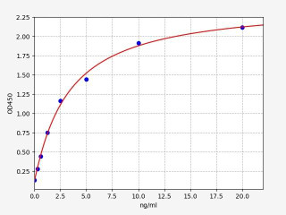
| Human CD68 / Macrosialin ELISA Kit | |
|---|---|
| ELISA Type | Sandwich |
| Sensitivity | 0.188ng/ml |
| Range | 0.313-20ng/ml |

| Human ITGAM / CD11b ELISA Kit | |
|---|---|
| ELISA Type | Sandwich |
| Sensitivity | 0.188ng/ml |
| Range | 0.313-20ng/ml |

| Human TREM2 / Triggering receptor expressed on myeloid cells 2 ELISA Kit | |
|---|---|
| ELISA Type | Sandwich |
| Sensitivity | 46.875pg/ml |
| Range | 78.125-5000pg/ml |
Written by Lauryn McLoughlin
Lauryn McLoughlin completed her undergraduate degree in Neuroscience before completing her masters in Biotechnology at University College Dublin.
Additional Resources
Recent Posts
-
Tigatuzumab Biosimilar: Harnessing DR5 for Targeted Cancer Therapy
Tigatuzumab is a monoclonal antibody targeting death receptor 5 (DR5), a member of the …17th Dec 2025 -
Enavatuzumab Biosimilar: Advancing TWEAKR-Targeted Therapy in Cancer
Enavatuzumab is a monoclonal antibody targeting TWEAK receptor (TWEAKR, also known as …17th Dec 2025 -
Alemtuzumab Biosimilar: Advancing CD52-Targeted Therapy
Alemtuzumab is a monoclonal antibody targeting CD52, a glycoprotein highly expressed o …17th Dec 2025

