The AP1M1 Antibody (CAB10129) is a high-quality antibody developed for reliable detection and analysis of target proteins. This antibody, produced in rabbits, has high specificity for human samples and is suitable for use in Western blot applications. By targeting AP1M1, researchers can analyze protein localization and trafficking mechanisms in various cell types, providing insights into cellular processes such as receptor recycling and secretion.AP1M1, a subunit of the adaptor protein complex 1 (AP-1), plays a crucial role in sorting proteins into vesicles for transport between intracellular compartments.
This antibody is validated for use in WB, ELISA applications and has demonstrated reactivity against Human, Mouse, Rat samples.
Product Name:
AP1M1 Antibody
SKU:
CAB10129
Size:
20μL, 100μL
Reactivity:
Human, Mouse, Rat
Conjugate:
Unconjugated
Immunogen:
Recombinant protein (or fragment).This information is considered to be commercially sensitive.
The protein encoded by this gene is the medium chain of the trans-Golgi network clathrin-associated protein complex AP-1. The other components of this complex are beta-prime-adaptin, gamma-adaptin, and the small chain AP1S1. This complex is located at the Golgi vesicle and links clathrin to receptors in coated vesicles. These vesicles are involved in endocytosis and Golgi processing. Alternatively spliced transcript variants encoding distinct protein isoforms have been found for this gene.
Purification Method
Affinity purification
Gene ID
8907
RRID
AB_2757654
Buffer Information
Store at -20℃. Avoid freeze / thaw cycles. Buffer: PBS containing 50% glycerol, preserved with proclin300 or sodium azide, pH 7.3.
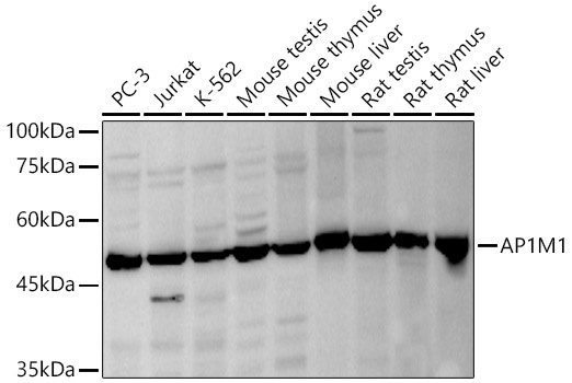
Western blot analysis of various lysates, using AP1M1 Rabbit pAb (CAB10129) at 1:1000 dilution. Secondary antibody: HRP-conjugated Goat anti-Rabbit IgG (H+L) (CABS014) at 1:10000 dilution. Lysates/proteins: 25μg per lane. Blocking buffer: 3% nonfat dry milk in TBST. Detection: ECL Basic Kit (AbGn00020). Exposure time: 30s.