The PAK3 Monoclonal Antibody (CAB3363) is a high-quality antibody developed for reliable detection and analysis of target proteins. This polyclonal antibody, produced in rabbits, is highly specific to human samples and has been validated for use in Western blot applications.PAK3 is a critical player in signal transduction pathways that control cell morphology and motility, making it a compelling target for investigations into cancer biology, neurodevelopmental disorders, and other conditions involving aberrant cell signaling.
This antibody is validated for use in WB, IHC-P, ELISA applications and has demonstrated reactivity against Mouse, Rat samples.
Product Name:
PAK3 Monoclonal Antibody
SKU:
CAB3363
Size:
20μL, 100μL
Reactivity:
Mouse, Rat
Clone Number:
ARC1955
Conjugate:
Unconjugated
Immunogen:
Synthetic peptide. This information is considered to be commercially sensitive.
The protein encoded by this gene is a serine-threonine kinase and forms an activated complex with GTP-bound RAS-like (P21), CDC2 and RAC1. This protein may be necessary for dendritic development and for the rapid cytoskeletal reorganization in dendritic spines associated with synaptic plasticity. Defects in this gene are the cause of a non-syndromic form of X-linked intellectual disability. Alternatively spliced transcript variants encoding different isoforms have been identified.
Purification Method
Affinity purification
Gene ID
5063
RRID
AB_2863043
Buffer Information
Store at -20℃. Avoid freeze / thaw cycles. Buffer: PBS containing 50% glycerol and 0.05% BSA, preserved with proclin300 or sodium azide, pH 7.3.
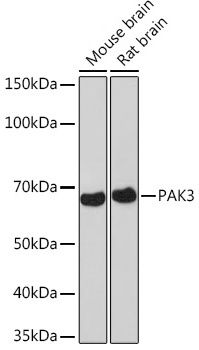
Western blot analysis of various lysates using PAK3 Rabbit mAb (CAB3363) at 1:1000 dilution. Secondary antibody: HRP-conjugated Goat anti-Rabbit IgG (H+L) (CABS014) at 1:10000 dilution. Lysates/proteins: 25μg per lane. Blocking buffer: 3% nonfat dry milk in TBST. Detection: ECL Basic Kit (AbGn00020). Exposure time: 3min.
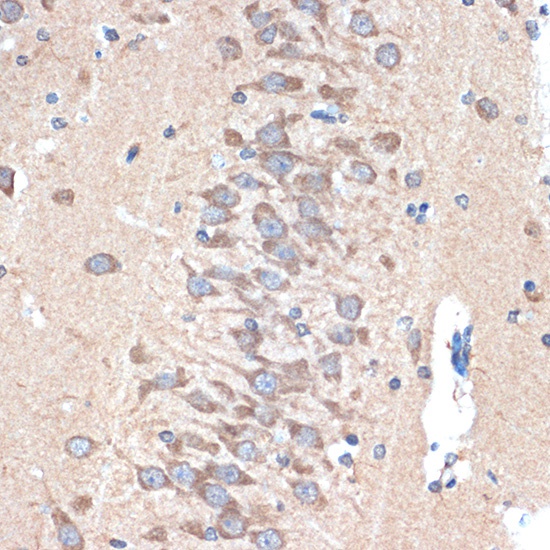
Immunohistochemistry analysis of paraffin-embedded Rat brain tissue using PAK3 Rabbit mAb (CAB3363) at dilution of 1:100 (40x lens). Microwave antigen retrieval performed with 0.01M Tris/EDTA Buffer (pH 9.0) prior to IHC staining.


![Anti-PAK3 [R05-7G2] Monoclonal Antibody (AGMB01887)](https://cdn11.bigcommerce.com/s-h68l9z2lnx/images/stencil/590x590/products/273176/676362/anti-pak3-r05-7g2-monoclonal-antibody-agmb01887__98435.1773029188.jpg?c=2 "Anti-PAK3 [R05-7G2] Monoclonal Antibody (AGMB01887)")


